Hyperbaric Oxygen Therapy (HBOT)
This is therapy that involves placing the patient into an airtight chamber and providing air/oxygen into it to provide an increased atmospheric pressure that then effects over 820 genes beginning after 30 minutes of being at this increased pressure (3.5-5 psi) providing a significant shift from inflammatory to anti-inflammatory gene expression. The increased pressure stimulates stem cells from the bone marrow to become activated and circulate in the blood. The increased oxygen in tissues also helps in preventing and fighting infections and in the healing of wounds and internal injured or degenerated tissues including the brain, spinal cord, nerves, blood vessels, pancreas, and other tissues that might have or be suffering from lower than normal oxygen levels or blood flow levels.
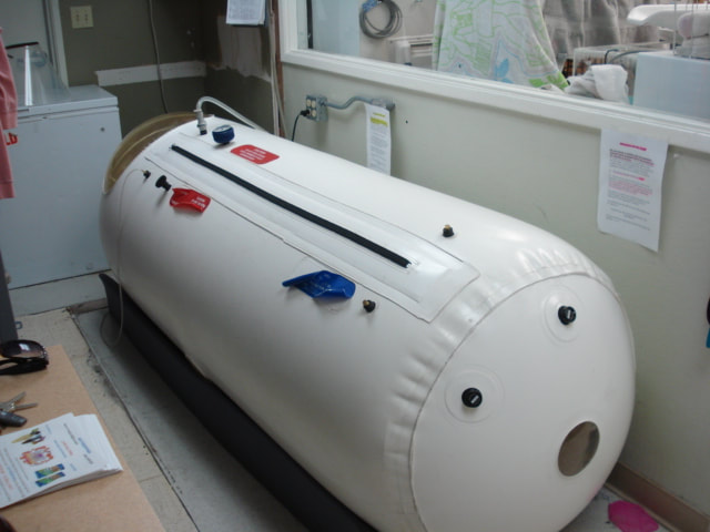
There are two types of hyperbaric oxygen chambers that Dr. Crowe uses - The one pictured above provides mild hyperbaric pressure and one that he can provide as its portable. He firmly believes in HBOT. He himself was treated with HBOT to prevent the loss of a leg following significant wounding and infection.
He is a member of the American College of Hyperbaric Medicine and is also a member of the International HyperbaricMedical Association. More information can be gained by going to www.hyperbaricmedicalassociation.org and then going to the research section. Please also go to the page "educational articles" in this website.
patients suffering from chronic neurological deficiencies
Recent studies, conducted at the Hyperbaric Center at Assaf Harofe Medical Center in patients suffering from chronic neurological deficiencies due to Stroke, Traumatic Brain Injury (TBI) or Anoxic Brain damage (ABD), have shown that HBOT can improve the symptoms and rejuvenate the damaged brain tissue. Treating Brain InjuriesAfter any brain injury there are several degrees of damage to the brain. The most severe is necrosis (the tissue is completely dead), and nothing can be done to help treat the necrotic area. However, surrounding the necrotic areas there are usually areas with metabolic dysfunctions, and this is where the hyperbaric oxygen can help. We can visualize these areas by metabolic imaging of the brain, we use SPECT analysis together with perfusion MRI+DTI, and in the location where is SPECT/MRI mismatch that is where the hyperbaric oxygen can help even years after the acute injury
STROKE The study of Stroke patients aimed to evaluate whether increasing the level of dissolved oxygen by Hyperbaric Oxygen Therapy (HBOT) could activate a regeneration of damaged brain tissue (neuroplasticity) in patients with chronic neurological deficiencies due to their Stroke. A prospective, randomized and controlled trial, including 74 patients was conducted (15 were excluded). All patients suffered a Stroke 6-36 months prior to inclusion and had at least one motor dysfunction. They all received two months of treatment, a total of 40 sessions, five days a week. The findings showed that the neurological functions and life quality of all the patients in the study had significantly improved following the HBOT sessions. The results of the SPECT imaging (CT) were well correlated with the clinical improvement of the patients, meaning that the imaging showed an improvement in blood supply and viability of damaged brain tissue. The results indicate that HBOT can lead to significant neurological improvements in post Stroke patients even years after the Stroke occurred. The observed clinical improvements imply that a regeneration of damaged brain cells can still be activated long after the acute brain insult.
In a second study with patients suffering from post Stroke symptoms, the researchers’ aim was to address the specific effects of HBOT on memory impairments after a Stroke at late chronic stages. The protocol included 40 to 60 daily session, five days per week, and memory cognitive tests were administered before and after HBOT Therapy. The results revealed significant improvements in all memory measures after HBOT treatments, and the clinical improvements were well correlated with improvements in brain metabolism. The study concluded that even though further research is needed, the results illustrate the potential of HBOT for improving memory impairments in post-Stroke patients, even years after the Stroke occurred.
Treating Traumatic Brain Injury (TBI)Traumatic Brain Injury (TBI) is defined as damage to the brain resulting from external mechanical force. TBI is the leading cause of death and disability in the US. Approximately 70-90% of the TBI cases are classified as mild, and up to 25% of them will not recover and suffer chronic neuro-cognitive impairment. Intensive therapy and rehabilitation programs are considered essential for maximizing quality of life but are often just partially successful. The main pathologies of these cases involve scattered brain injuries, which are hard to detect and locate. The Study that was conducted at the Hyperbaric Center at Assaf Harofe Medical Center tested the effectiveness of HBOT in improving brain function and quality of life in TBI patients suffering chronic neuro-cognitive impairments. The Study included 56 patients 1-5 years after an injury with prolonged post-concussion Syndrome (PCS). They received 40 HBOT treatments, five days a week. Significant improvements were demonstrated in cognitive function and “quality of life” following HBOT, and the brain imaging revealed an elevated brain activity correlating with the cognitive improvements. The study concluded that HBOT can induce a regeneration/rejuvenation of damaged brain tissue, leading to the repair of chronically impaired brain functions and improved quality of life in TBI patients with prolonged post-concussion Syndrome even years after the injury occurred.
Rehabilitation UnitThe Neurological Rehabilitation Unit is a division of the Sagol Center, integrating several rehabilitation disciplines in one location (neuropsychology, physiotherapy, and speech-therapy). The biological intervention (HBOT) together with intensive multidisciplinary rehabilitation introduces a new promising venue for achieving a fast and potent recovery process, enabling the patients to regain their normal lives.
Patients treated in the Unit initially undergo a baseline interdisciplinary assessment process, evaluating their motor, cognitive, emotional and speech impairments. Based on the results, a specific rehabilitation program is designed for each patient that includes individual cognitive rehabilitation treatments, group cognitive rehabilitation sessions, computerized cognitive training, physiotherapy, speech-therapy and acupuncture.
Rehabilitation is short and intensive requiring highly motivated patients. The Unit is suitable for adult patients suffering from post concussion syndrome, stroke, anoxic brain damage, traumatic brain injury, mild cognitive impairment or dementia.Anoxic Brain Damage (ABD)Cognitive impairment may occur in 42-50% of cardiac arrest survivors. An additional pilot study conducted at the Sagol Hyperbaric Center at Assaf Harofe Medical Center, where patients were treated with Hyperbaric Oxygen (HBOT) 0.5-7.5 (mean 2.6+_0.6 years)after the cardiac arrest, Even though HBOT was started at the late chronic phase, it induced significant cognitive improvements in all of the patients. The clinical improvements were well documented by neuro-cognitive tests and correlated with improved ability to perform the activities of daily living and quality of life. The most significant measurable improvements were in executive functions, attention and memory. The clinical improvement correlated with metabolic improvement of the injured brain tissue as was well visualized by brain metabolic imaginRead the Full Articles
Hyperbaric Oxygen Induces Late Neuroplasticity in Post Stroke Patients - Randomized, Prospective Trial
Recovery after stroke correlates with non-active (stunned) brain regions, which may persist for years. The current study aimed to evaluate whether increasing the level of dissolved oxygen by Hyperbaric Oxygen Therapy (HBOT) could activate neuroplasticity in patients with chronic neurologic deficiencies due to stroke.
Methods and FindingsA prospective, randomized, controlled trial including 74 patients (15 were excluded). All participants suffered a stroke 6–36 months prior to inclusion and had at least one motor dysfunction. After inclusion, patients were randomly assigned to "treated" or "cross" groups. Brain activity was assessed by SPECT imaging; neurologic functions were evaluated by NIHSS, ADL, and life quality. Patients in the treated group were evaluated twice: at baseline and after 40 HBOT sessions. Patients in the cross group were evaluated three times: at baseline, after a 2-month control period of no treatment, and after subsequent 2-months of 40 HBOT sessions. HBOT protocol: Two months of 40 sessions (5 days/week), 90 minutes each, 100% oxygen at 2 ATA. We found that the neurological functions and life quality of all patients in both groups were significantly improved following the HBOT sessions while no improvement was found during the control period of the patients in the cross group. Results of SPECT imaging were well correlated with clinical improvement. Elevated brain activity was detected mostly in regions of live cells (as confirmed by CT) with low activity (based on SPECT) – regions of noticeable discrepancy between anatomy and physiology.
ConclusionsThe results indicate that HBOT can lead to significant neurological improvements in post stroke patients even at chronic late stages. The observed clinical improvements imply that neuroplasticity can still be activated long after damage onset in regions where there is a brain SPECT/CT (anatomy/physiology) mismatch.
IntroductionIntensive functional therapy and rehabilitation programs for post stroke patients are considered essential for maximizing the patients' quality of life [1], [2]. Unfortunately, these programs are often just partially successful, and additional therapeutic approaches towards metabolic recovery of affected cerebral tissues are called for. While a considerable amount of preclinical research supports the use of hyperbaric oxygen therapy (HBOT) for post-stroke damaged brain tissue, so far, only 5 articles reported controlled clinical trials of HBOT for stroke patients. These studies, in which the treatment started during the early-acute phase immediately after stroke, yielded non conclusive and somewhat contradicting results [3], [4], [5], [6], [7]. In contrast, a recent phase-I study evaluating the effect of HBOT on chronic neurological deficiencies (due to traumatic brain injury) revealed promising results [8]. However, to date the effects of HBOT on neurological deficiencies due to stroke during the late-chronic phase (the focus of the current report) have not yet been investigated in a prospective randomized trial.
Years of clinical experience revealed that the dramatic spontaneous recovery from stroke occurs mainly within the first 30 days, though moderate and severe stroke survivors continue to improve for at least 90 days [9]. Most of the recovery involves brain regions rendered dysfunctional, but not dead [10]. Accumulated data from visualizations of these non-active (stunned) regions indicates that they may persist alive but dysfunctional for months, even years, after the acute injury [11], [12], [13]. It was proposed that the oxygen supply to these under-active neurons was low due to stroke damage to blood vessels in these regions, leading to oxygen deficiency, anaerobic metabolism and ATP depletion [14], [15]. The decreased oxygen level not only causes reduction in the neuronal activity but also prevents angiogenesis to replace the stroke-damaged blood vessels and the generation of new synaptic connections. Since 1 cm3 of normal brain tissue contains about 1 km of blood vessels, high oxygen supply is essential for repair of the stunned regions. Indeed, as has been demonstrated by previous studies, an increase in dissolved oxygen has several beneficial effects in damaged brain tissues [13], [16], [17], [18], [19], [20]. Transport of oxygen to glial mitochondria, the main sites of oxygen utilization, follows oxygen release from erythrocytes into the plasma and then diffusion of the blood-dissolved oxygen across the Blood-Brain Barrier (BBB). Breathing oxygen under hyperbaric conditions has been shown to be a potent means of increasing arterial oxygen tension and consequently the brain oxygen tension [20], [21], [22]. For example, at 2ATA (atmospheres absolute), the plasma O2 partial pressure rises above 1,110 mmHg. Hence, it is reasonable to expect that HBOT can be an efficient (and clinically feasible) method for increasing tissue/cellular oxygenation and thus effectively evoking neuroplasticity in the chronically non-active areas during the late post-stroke phase.
Many physiological pathways, each with a different characteristic time, are spontaneously activated following the onset of stroke. Therefore, a challenging question to be addressed considers the optimal time lapse after stroke to start the HBOT procedure. It should also be kept in mind that signals and chemical cues associated with cell death during the acute stage of stroke might, in fact, promote repair during recovery [23] and can be negatively affected by premature application of HBOT. Unlike the case of preclinical animal studies, in clinical practice it is not feasible to apply the HBOT immediately at the stroke onset. Thus, HBOT procedure can practically begin either at the degenerative or at the regenerative stage. One can assume that any added energy during the degenerative stage could further increase the unwanted, post-injury damage. On the other hand, elevated oxygen supply during the regenerative stage would supply the energy needs for the innate brain repair processes. The differences in the time lapse between stroke onset and HBOT application in previous studies are likely to be the reason for the contradictive results obtained for HBOT application during the acute phase after stroke [3], [4], [5], [6], [7]. The aim of the current study was to evaluate the effects of HBOT started at the late-chronic phase after the acute stroke.
MethodsThe study was performed as a prospective, randomized, controlled, two-group trial. The population included patients of ages 18 years or older, who had either ischemic or hemorrhagic stroke 6–36 months prior to their inclusion. All patients had to have at least one motor dysfunction. Exclusions were based on chest pathology incompatible with HBOT, inner ear disease, claustrophobia and inability to sign informed consent. Additional exclusions were based on dynamic neurologic improvements during the last month (based either on objective measurements by external evaluator or on subjective statement by the patients). Smoking was not allowed during the study. All patients signed written informed consent; the protocol was approved by the local Helsinki committee. The study was conducted in the hyperbaric and research units of Assaf-Harofeh Medical Center, Israel.
Protocol and End PointsAfter signing an informed consent form, the patients were invited for baseline evaluations. Included patients were randomized into two groups (1∶1 randomization): a treated group and a cross group. The neurologic functions as evaluated by National Institutes of Health Stroke Scale (NIHSS) [24], [25], ability to perform activities of daily living (ADL) [26], and brain metabolism as visualized SPECT were the primary endpoints of the study. The secondary end point of the study included Quality of life evaluation. The patients in the treated group were evaluated twice – at baseline and after 2 months of HBOT treatment. Patients in the cross group were evaluated three times: baseline, after 2 months control period of no treatment, and after consequent 2 months of HBOT sessions (Figure 1). The post-HBOT neurological evaluations as well as the SPECT scans were performed more than 1 week (1–3 weeks) after the end of the HBOT protocol. The following HBOT protocol was practiced: 40 daily sessions, 5 days/week, 90 minutes each, 100% oxygen at 2ATA. The detailed clinical study protocol (Protocol S1), randomization and placebo consideration (Text S1), copy of the informed consent (Form S1), as well as CONSORT 2010 checklist of information (Checklist S1) are attached as supporting information.
STROKE The study of Stroke patients aimed to evaluate whether increasing the level of dissolved oxygen by Hyperbaric Oxygen Therapy (HBOT) could activate a regeneration of damaged brain tissue (neuroplasticity) in patients with chronic neurological deficiencies due to their Stroke. A prospective, randomized and controlled trial, including 74 patients was conducted (15 were excluded). All patients suffered a Stroke 6-36 months prior to inclusion and had at least one motor dysfunction. They all received two months of treatment, a total of 40 sessions, five days a week. The findings showed that the neurological functions and life quality of all the patients in the study had significantly improved following the HBOT sessions. The results of the SPECT imaging (CT) were well correlated with the clinical improvement of the patients, meaning that the imaging showed an improvement in blood supply and viability of damaged brain tissue. The results indicate that HBOT can lead to significant neurological improvements in post Stroke patients even years after the Stroke occurred. The observed clinical improvements imply that a regeneration of damaged brain cells can still be activated long after the acute brain insult.
In a second study with patients suffering from post Stroke symptoms, the researchers’ aim was to address the specific effects of HBOT on memory impairments after a Stroke at late chronic stages. The protocol included 40 to 60 daily session, five days per week, and memory cognitive tests were administered before and after HBOT Therapy. The results revealed significant improvements in all memory measures after HBOT treatments, and the clinical improvements were well correlated with improvements in brain metabolism. The study concluded that even though further research is needed, the results illustrate the potential of HBOT for improving memory impairments in post-Stroke patients, even years after the Stroke occurred.
Treating Traumatic Brain Injury (TBI)Traumatic Brain Injury (TBI) is defined as damage to the brain resulting from external mechanical force. TBI is the leading cause of death and disability in the US. Approximately 70-90% of the TBI cases are classified as mild, and up to 25% of them will not recover and suffer chronic neuro-cognitive impairment. Intensive therapy and rehabilitation programs are considered essential for maximizing quality of life but are often just partially successful. The main pathologies of these cases involve scattered brain injuries, which are hard to detect and locate. The Study that was conducted at the Hyperbaric Center at Assaf Harofe Medical Center tested the effectiveness of HBOT in improving brain function and quality of life in TBI patients suffering chronic neuro-cognitive impairments. The Study included 56 patients 1-5 years after an injury with prolonged post-concussion Syndrome (PCS). They received 40 HBOT treatments, five days a week. Significant improvements were demonstrated in cognitive function and “quality of life” following HBOT, and the brain imaging revealed an elevated brain activity correlating with the cognitive improvements. The study concluded that HBOT can induce a regeneration/rejuvenation of damaged brain tissue, leading to the repair of chronically impaired brain functions and improved quality of life in TBI patients with prolonged post-concussion Syndrome even years after the injury occurred.
Rehabilitation UnitThe Neurological Rehabilitation Unit is a division of the Sagol Center, integrating several rehabilitation disciplines in one location (neuropsychology, physiotherapy, and speech-therapy). The biological intervention (HBOT) together with intensive multidisciplinary rehabilitation introduces a new promising venue for achieving a fast and potent recovery process, enabling the patients to regain their normal lives.
Patients treated in the Unit initially undergo a baseline interdisciplinary assessment process, evaluating their motor, cognitive, emotional and speech impairments. Based on the results, a specific rehabilitation program is designed for each patient that includes individual cognitive rehabilitation treatments, group cognitive rehabilitation sessions, computerized cognitive training, physiotherapy, speech-therapy and acupuncture.
Rehabilitation is short and intensive requiring highly motivated patients. The Unit is suitable for adult patients suffering from post concussion syndrome, stroke, anoxic brain damage, traumatic brain injury, mild cognitive impairment or dementia.Anoxic Brain Damage (ABD)Cognitive impairment may occur in 42-50% of cardiac arrest survivors. An additional pilot study conducted at the Sagol Hyperbaric Center at Assaf Harofe Medical Center, where patients were treated with Hyperbaric Oxygen (HBOT) 0.5-7.5 (mean 2.6+_0.6 years)after the cardiac arrest, Even though HBOT was started at the late chronic phase, it induced significant cognitive improvements in all of the patients. The clinical improvements were well documented by neuro-cognitive tests and correlated with improved ability to perform the activities of daily living and quality of life. The most significant measurable improvements were in executive functions, attention and memory. The clinical improvement correlated with metabolic improvement of the injured brain tissue as was well visualized by brain metabolic imaginRead the Full Articles
- Hyperbaric Oxygen Induces Late Neuroplasticity in Post Stroke Patients - Click Here!
- Hyperbaric Oxygen Therapy Can Improve Post Concussion Syndrome Years after Mild Traumatic Brain Injury - Click Here!
- Improvement of Memory Impairments in Post stroke Patients by Hyperbaric Oxygen Therapy - Click here!
- How and why hyperbaric oxygen therapy can bring new hope for children suffering from cerebral palsy - Click Here!
- Intensive rehabilitation combined with HBO2 therapy in children with cerebral palsy: A controlled longitudinal study - Click Here!
- Hyperbaric Oxygen Therapy Can Diminish Fibromyalgia Syndrome – Prospective Clinical Trial - Click Here!
- Hyperbaric oxygen can induce angiogenesis in post concussion patients - Click Here!
- Oxygen - a limiting factor for brain recovery - Click Here!
- Reflection on the neurotherapeutic effects of hyperbaric oxygen - Click Here!
Hyperbaric Oxygen Induces Late Neuroplasticity in Post Stroke Patients - Randomized, Prospective Trial
- Shai Efrati ,
- Gregori Fishlev,
- Yair Bechor,
- Olga Volkov,
- Jacob Bergan,
- Kostantin Kliakhandler,
- Izhak Kamiager,
- Nachum Gal,
- Mony Friedman,
- Eshel Ben-Jacob,
- Haim Golan
- Published: January 15, 2013
- http://dx.doi.org/10.1371/journal.pone.0053716
Recovery after stroke correlates with non-active (stunned) brain regions, which may persist for years. The current study aimed to evaluate whether increasing the level of dissolved oxygen by Hyperbaric Oxygen Therapy (HBOT) could activate neuroplasticity in patients with chronic neurologic deficiencies due to stroke.
Methods and FindingsA prospective, randomized, controlled trial including 74 patients (15 were excluded). All participants suffered a stroke 6–36 months prior to inclusion and had at least one motor dysfunction. After inclusion, patients were randomly assigned to "treated" or "cross" groups. Brain activity was assessed by SPECT imaging; neurologic functions were evaluated by NIHSS, ADL, and life quality. Patients in the treated group were evaluated twice: at baseline and after 40 HBOT sessions. Patients in the cross group were evaluated three times: at baseline, after a 2-month control period of no treatment, and after subsequent 2-months of 40 HBOT sessions. HBOT protocol: Two months of 40 sessions (5 days/week), 90 minutes each, 100% oxygen at 2 ATA. We found that the neurological functions and life quality of all patients in both groups were significantly improved following the HBOT sessions while no improvement was found during the control period of the patients in the cross group. Results of SPECT imaging were well correlated with clinical improvement. Elevated brain activity was detected mostly in regions of live cells (as confirmed by CT) with low activity (based on SPECT) – regions of noticeable discrepancy between anatomy and physiology.
ConclusionsThe results indicate that HBOT can lead to significant neurological improvements in post stroke patients even at chronic late stages. The observed clinical improvements imply that neuroplasticity can still be activated long after damage onset in regions where there is a brain SPECT/CT (anatomy/physiology) mismatch.
IntroductionIntensive functional therapy and rehabilitation programs for post stroke patients are considered essential for maximizing the patients' quality of life [1], [2]. Unfortunately, these programs are often just partially successful, and additional therapeutic approaches towards metabolic recovery of affected cerebral tissues are called for. While a considerable amount of preclinical research supports the use of hyperbaric oxygen therapy (HBOT) for post-stroke damaged brain tissue, so far, only 5 articles reported controlled clinical trials of HBOT for stroke patients. These studies, in which the treatment started during the early-acute phase immediately after stroke, yielded non conclusive and somewhat contradicting results [3], [4], [5], [6], [7]. In contrast, a recent phase-I study evaluating the effect of HBOT on chronic neurological deficiencies (due to traumatic brain injury) revealed promising results [8]. However, to date the effects of HBOT on neurological deficiencies due to stroke during the late-chronic phase (the focus of the current report) have not yet been investigated in a prospective randomized trial.
Years of clinical experience revealed that the dramatic spontaneous recovery from stroke occurs mainly within the first 30 days, though moderate and severe stroke survivors continue to improve for at least 90 days [9]. Most of the recovery involves brain regions rendered dysfunctional, but not dead [10]. Accumulated data from visualizations of these non-active (stunned) regions indicates that they may persist alive but dysfunctional for months, even years, after the acute injury [11], [12], [13]. It was proposed that the oxygen supply to these under-active neurons was low due to stroke damage to blood vessels in these regions, leading to oxygen deficiency, anaerobic metabolism and ATP depletion [14], [15]. The decreased oxygen level not only causes reduction in the neuronal activity but also prevents angiogenesis to replace the stroke-damaged blood vessels and the generation of new synaptic connections. Since 1 cm3 of normal brain tissue contains about 1 km of blood vessels, high oxygen supply is essential for repair of the stunned regions. Indeed, as has been demonstrated by previous studies, an increase in dissolved oxygen has several beneficial effects in damaged brain tissues [13], [16], [17], [18], [19], [20]. Transport of oxygen to glial mitochondria, the main sites of oxygen utilization, follows oxygen release from erythrocytes into the plasma and then diffusion of the blood-dissolved oxygen across the Blood-Brain Barrier (BBB). Breathing oxygen under hyperbaric conditions has been shown to be a potent means of increasing arterial oxygen tension and consequently the brain oxygen tension [20], [21], [22]. For example, at 2ATA (atmospheres absolute), the plasma O2 partial pressure rises above 1,110 mmHg. Hence, it is reasonable to expect that HBOT can be an efficient (and clinically feasible) method for increasing tissue/cellular oxygenation and thus effectively evoking neuroplasticity in the chronically non-active areas during the late post-stroke phase.
Many physiological pathways, each with a different characteristic time, are spontaneously activated following the onset of stroke. Therefore, a challenging question to be addressed considers the optimal time lapse after stroke to start the HBOT procedure. It should also be kept in mind that signals and chemical cues associated with cell death during the acute stage of stroke might, in fact, promote repair during recovery [23] and can be negatively affected by premature application of HBOT. Unlike the case of preclinical animal studies, in clinical practice it is not feasible to apply the HBOT immediately at the stroke onset. Thus, HBOT procedure can practically begin either at the degenerative or at the regenerative stage. One can assume that any added energy during the degenerative stage could further increase the unwanted, post-injury damage. On the other hand, elevated oxygen supply during the regenerative stage would supply the energy needs for the innate brain repair processes. The differences in the time lapse between stroke onset and HBOT application in previous studies are likely to be the reason for the contradictive results obtained for HBOT application during the acute phase after stroke [3], [4], [5], [6], [7]. The aim of the current study was to evaluate the effects of HBOT started at the late-chronic phase after the acute stroke.
MethodsThe study was performed as a prospective, randomized, controlled, two-group trial. The population included patients of ages 18 years or older, who had either ischemic or hemorrhagic stroke 6–36 months prior to their inclusion. All patients had to have at least one motor dysfunction. Exclusions were based on chest pathology incompatible with HBOT, inner ear disease, claustrophobia and inability to sign informed consent. Additional exclusions were based on dynamic neurologic improvements during the last month (based either on objective measurements by external evaluator or on subjective statement by the patients). Smoking was not allowed during the study. All patients signed written informed consent; the protocol was approved by the local Helsinki committee. The study was conducted in the hyperbaric and research units of Assaf-Harofeh Medical Center, Israel.
Protocol and End PointsAfter signing an informed consent form, the patients were invited for baseline evaluations. Included patients were randomized into two groups (1∶1 randomization): a treated group and a cross group. The neurologic functions as evaluated by National Institutes of Health Stroke Scale (NIHSS) [24], [25], ability to perform activities of daily living (ADL) [26], and brain metabolism as visualized SPECT were the primary endpoints of the study. The secondary end point of the study included Quality of life evaluation. The patients in the treated group were evaluated twice – at baseline and after 2 months of HBOT treatment. Patients in the cross group were evaluated three times: baseline, after 2 months control period of no treatment, and after consequent 2 months of HBOT sessions (Figure 1). The post-HBOT neurological evaluations as well as the SPECT scans were performed more than 1 week (1–3 weeks) after the end of the HBOT protocol. The following HBOT protocol was practiced: 40 daily sessions, 5 days/week, 90 minutes each, 100% oxygen at 2ATA. The detailed clinical study protocol (Protocol S1), randomization and placebo consideration (Text S1), copy of the informed consent (Form S1), as well as CONSORT 2010 checklist of information (Checklist S1) are attached as supporting information.
Hyperbaric oxygen articles
Overview: Hyperbaric Oxygen Therapy (HBOT)
This is therapy that involves placing the patient into an airtight chamber and providing air/oxygen into it to provide an increased atmospheric pressure that then effects over 820 genes beginning after 30 minutes of being at this increased pressure (3.5-5 psi) providing a significant shift from inflammatory to anti-inflammatory gene expression. The increased pressure stimulates stem cells from the bone marrow to become activated and circulate in the blood. The increased oxygen in tissues also helps in preventing and fighting infections and in the healing of wounds and internal injured or degenerated tissues including the brain, spinal cord, nerves, blood vessels, pancreas, and other tissues that might have or be suffering from lower than normal oxygen levels or blood flow levels.
There are two types of hyperbaric oxygen chambers that Dr. Crowe uses - The one pictured above provides mild hyperbaric pressure and one that he can provide as its portable. He firmly believes in HBOT. He himself was treated with HBOT to prevent the loss of a leg following significant wounding and infection. He is a member of the American College of Hyperbaric Medicine and is also a member of the International HyperbaricMedical Association. More information can be gained by going to www.hyperbaricmedicalassociation.org and then going to the research section. Please also go to the page "educational articles" in this website.
Hyperbaric oxygen articles
Evidence Report/Technology Assessment: Number 85
Under its Evidence-based Practice Program, the Agency for Healthcare Research and Quality (AHRQ) is developing scientific information for other agencies and organizations on which to base clinical guidelines, performance measures, and other quality improvement tools. Contractor institutions review all relevant scientific literature on assigned clinical care topics and produce evidence reports and technology assessments, conduct research on methodologies and the effectiveness of their implementation, and participate in technical assistance activities.
Overview
Hyperbaric oxygen therapy (HBOT) is the inhalation of 100 percent oxygen inside a hyperbaric chamber that is pressurized to greater than 1 atmosphere (atm). HBOT causes both mechanical and physiologic effects by inducing a state of increased pressure and hyperoxia. HBOT is typically administered at 1 to 3 atm. While the duration of an HBOT session is typically 90 to 120 minutes, the duration, frequency, and cumulative number of sessions have not been standardized.
HBOT is administered in two primary ways, using a monoplace chamber or a multiplace chamber. The monoplace chamber is the less-costly option for initial setup and operation but provides less opportunity for patient interaction while in the chamber. Multiplace chambers allow medical personnel to work in the chamber and care for acute patients to some extent. The entire multiplace chamber is pressurized, so medical personnel may require a controlled decompression, depending on how long they were exposed to the hyperbaric air environment.
The purpose of this report is to provide a guide to the strengths and limitations of the evidence about the use of HBOT to treat patients who have brain injury, cerebral palsy, and stroke.
Brain injury can be caused by an external physical force (also known as traumatic brain injury, or TBI); rapid acceleration or deceleration of the head; bleeding within or around the brain; lack of sufficient oxygen to the brain; or toxic substances passing through the blood-brain barrier. Brain injury results in temporary or permanent impairment of cognitive, emotional, and/or physical functioning. Cerebral palsy refers to a motor deficit that usually manifests itself by 2 years of age and is secondary to an abnormality of at least the part of the brain that relates to motor function. Stroke refers to a sudden interruption of the blood supply to the brain, usually caused by a blocked artery or a ruptured blood vessel, leading to an interruption of homeostasis of cells, and symptoms such as loss of speech and loss of motor function.
While these conditions have different etiologies, prognostic factors, and outcomes, they also have important similarities. Each condition represents a broad spectrum, from barely perceptible or mild disabilities to devastating ones. All three are characterized by acute and chronic phases and by changes over time in the type and degree of disability. Another similarity is that the outcome of conventional treatment is often unsatisfactory. For brain injury in particular, there is a strong sense that conventional treatment has made little impact on outcomes.
Predicting the outcome of brain injury, cerebral palsy, and stroke is difficult. Prognostic instruments, such as the Glasgow Coma Scale (GCS) for brain injury, are not precise enough to reliably predict an individual patient's mortality and long-term functional status. Various prognostic criteria for the cerebral palsy patient's function have been developed over the years. For example, if a patient is not sitting independently when placed by age 2, then one can predict with approximately 95 percent confidence that he/she never will be able to walk. However, it is not possible to predict precisely when an individual patient is likely to acquire a particular ability, such as smiling, recognizing other individuals, or saying or understanding a new word.
Mortality and morbidity from a stroke are related to older age, history of myocardial infarction, cardiac arrhythmias, diabetes mellitus, and the number of stroke deficits. Functional recovery is dependent on numerous variables, including age, neurologic deficit, comorbidities, psychosocial factors, educational level, vocational status, and characteristics of the stroke survivor's environment.
The report focuses on the quality and consistency of studies reporting clinical outcomes of the use of HBOT in humans who have brain injury, cerebral palsy, or stroke. This information can be used to help providers counsel patients who use this therapy and to identify future research needs.
Reporting the Evidence
This review addresses the following questions:
Does HBOT improve mortality and morbidity in patients who have traumatic brain injury or nontraumatic brain injury, such as anoxic ischemic encephalopathy?
Does HBOT improve functional outcomes in patients who have cerebral palsy? (Examples of improved functional outcomes are decreased spasticity, improved speech, increased alertness, increased cognitive abilities, and improved visual functioning.)
Does HBOT improve mortality and morbidity in patients who have suffered a stroke?
What are the adverse effects of using HBOT in these conditions?
To identify the patient groups, interventions, and outcomes that should be included in the review, we read background material from diverse sources including textbooks, government reports, proceedings of scientific meetings, and Web sites. We also conducted focus groups and interviews to improve our understanding of the clinical logic underlying the rationale for the use of HBOT. In the focus groups, we identified outcomes of treatment with HBOT that are important to patients, caregivers, and clinicians and examined whether patients, caregivers, and clinicians who have experience with HBOT value certain outcomes differently from those who have not used HBOT. A broader goal of the focus groups was to better understand the disagreement between supporters and non-supporters of HBOT.
The following interventions, populations, outcomes, and study design criteria were used to formulate the literature search strategy and to assess eligibility of studies.
Intervention. Hyperbaric oxygen therapy: any treatment using 100 percent oxygen supplied to a patient inside a hyperbaric chamber that is pressurized to greater than 1 atm.
Population. Patients with:
Brain injury from any cause and in any stage (acute, subacute, or chronic).
Cerebral palsy of any etiology.
Thrombotic stroke.
Outcomes. We sought articles reporting any clinical endpoint. We focused on health outcomes, including mortality and functional changes that a patient would experience, rather than intermediate outcomes. Intermediate outcomes include physiologic measures, such as intracranial pressure, cerebrospinal fluid lactate levels, or changes in cerebral blood flow, or results of imaging studies. Some clinical measures, such as neuropsychiatric and cognitive tests, are also intermediate measures. We did not assume that any of these intermediate measures of the effect of HBOT on patients with brain injury, cerebral palsy, or stroke was proven to be an indicator of the long-term outcome. Instead, in reviewing articles for inclusion in this report, we were particularly interested in studies that reported both intermediate measures and health outcomes, to assess the strength of evidence about their correlation.
Design. We included original studies of human subjects that reported original data (no reviews). All study designs except for case reports and small case series were eligible for inclusion. Before-after or time-series studies with no independent control group were included if a) five or more cases were reported, and b) outcome measures were reported for both the pre- and post-HBOT period.
Methodology
Technical Expert Advisory Group (TEAG)
We identified technical experts to assist us in formulating the research questions and identifying relevant databases for the literature search. The expert panelists included a neurologist specializing in stroke, a neurosurgeon specializing in severe brain injury, a pediatric neurologist with expertise in treating patients with cerebral palsy, and a physician with an HBOT practice. Throughout the project period, we consulted individual members of the TEAG on issues that arose in the course of identifying and reviewing the literature.
Literature Search, Study Selection, and Data Extraction
We searched a broad range of databases to identify published and unpublished studies of the effectiveness and harms of HBOT in patients with brain injury, cerebral palsy, and stroke. Each database was searched from its starting date to March 2001. The databases searched were:
MEDLINE®. HealthSTAR (Health Service Technology, Administration and Research).
CINAHL® (Cumulative Index to Nursing & Allied Health).
Cochrane Database of Systematic Reviews.
Cochrane Controlled Trials Register.
DARE (Database of Abstracts of Reviews of Effectiveness). AltHealthWatch.
MANTIS™ (Manual, Alternative and Natural Therapy).
Health Technology Assessment Database.
TEAG members identified the following additional databases as potential sources of other material that may not be indexed in other electronic databases:
The Undersea & Hyperbaric Medical Society: a large bibliographic database.
The Database of Randomized Controlled Trials In Hyperbaric Medicine.
European Underwater and Baromedical Society.
International Congress on Hyperbaric Medicine.
National Baromedical Services, Inc.
Update literature searching of the electronic databases MEDLINE®, PreMEDLINE®, EMBASE, CINAHL®, the Cochrane Library, and the Health Technology Assessment Database was completed on February 26, 2002, using the same search strategy as used for the initial searches. Eight additional references submitted by a peer reviewer were added in May 2003. Finally, a supplemental search of MEDLINE®, PreMEDLINE®, EMBASE, and CINAHL® was conducted in July 2003.
The references of all included papers were hand searched. In addition, two reviewers independently conducted hand searches of the references from the Textbook of Hyperbaric Medicine.1 One TEAG member provided articles and meeting abstracts from his personal library.
Two reviewers independently assessed each title and abstract located through the literature searches for relevance to the review, based on the intervention, population, outcome, and study design criteria. The full-text articles, reports, or meeting abstracts that met the criteria listed above were retrieved and reviewed independently by two reviewers who reapplied the eligibility criteria. Disagreements were resolved through consensus.
Extraction of data from studies was performed by one reviewer and checked by a second reviewer. Disagreements were resolved through consensus.
Internal and External Validity and Quality Rating
The quality of all trials in the review was assessed using a list of items indicating components of internal validity. We modified the standard checklists to address issues of particular importance in studies of HBOT. For randomized controlled trials (RCTs) and nonrandomized controlled trials (NRCTs), the items assessed for internal validity were: randomization/allocation concealment, baseline comparability of groups, timing of baseline measures, intervention, outcome measures, timing of followup measurements (long enough to assess effects), loss to followup, handling of dropouts or missing data, masking, statistical analysis (if any), and general reviewer comments.
For the observational studies, items assessed for internal validity were exposure measurement (whether all subjects were given the same HBOT treatment), other interventions, differences in baseline factors among the groups of subjects compared (if a comparison group was included), discussion of or control for potential confounding, masking, evidence of stable baseline, timing of baseline survey, timing of followup measures, outcome measures used, and general comments of the reviewer.
Each study was then assigned an overall rating (good, fair or poor) according to the US Preventive Services Task Force method:
Good: Comparable groups assembled initially (adequate randomization and concealment, and potential confounders distributed equally among groups) and maintained throughout the study; followup at least 80 percent; reliable and valid measurement instruments applied equally to the groups; outcome assessment masked; interventions defined clearly; all important outcomes considered; appropriate attention to confounders in analysis; for RCTs, intention-to-treat analysis.
Fair: Generally comparable groups assembled initially (inadequate or unstated randomization and concealment methods) but some question remains whether some (although not major) differences occurred with followup; measurement instruments acceptable (although not the best) and generally applied equally; outcome assessment masked; some, but not all, important outcomes considered; appropriate attention to some, but not all, potential confounders; for RCTs, intention-to-treat analysis.
Poor: Groups assembled initially not close to being comparable or not maintained throughout the study; measurement instruments unreliable or invalid or not applied equally among groups; outcome assessment not masked; key confounders given little or no attention; for RCTs, no intention-to-treat analysis.
For each study, the reviewer's assessment of external validity is given, including an assessment of the evidence that the study population reflects the underlying patient population (age-range, co-morbidities, co-interventions, etc.). External validity indicates the applicability of the results of the study to clinical practice. For example, if the study recruited a narrowly defined group of patients, the results may not be generalizable to a broader spectrum of patients. A study can have high internal validity but low external validity. There are no well-defined criteria for assessing external validity, and clinicians must assess the applicability of the results to the patient population for which the intervention is intended.
1 Jain K, editor. Textbook of hyperbaric medicine. 3rd rev. ed. Kirkland, WA: Hogrefe & Huber Publishers, Inc; 1999
Findings: Brain Injury
For traumatic brain injury, one randomized trial provided fair evidence that HBOT might reduce mortality or the duration of coma in severely injured TBI (traumatic brain injuries) patients. However, in this trial, HBOT also increased the chance of a poor functional outcome. A second fair quality randomized trial found no difference in mortality or morbidity overall, but a significant reduction in mortality in one subgroup. Therefore, they provide insufficient evidence to determine whether the benefits of HBOT outweigh the potential harms.
The quality of the controlled trials was fair, meaning that deficiencies in the design add to uncertainty about the validity of results.
Due to flaws in design or small size, the observational studies of HBOT in TBI do not establish a clear, consistent relationship between physiologic changes after HBOT sessions and measures of clinical improvement.
The evidence for use of HBOT in other types of brain injury is inconclusive. No good- or fair-quality studies were found.
Cerebral Palsy
There is insufficient evidence to determine whether the use of HBOT improves functional outcomes in children with cerebral palsy. The results of the only truly randomized trial were difficult to interpret because of the use of pressurized room air in the control group. As both groups improved, the benefit of pressurized air and of HBOT at 1.3 to 1.5 atm should both be examined in future studies.
The only other controlled study compared HBOT treatments with 1.5 atm to delaying treatment for 6 months. As in the placebo-controlled study, significant improvements were seen, but there was not a significant difference between groups.
Two fair-quality uncontrolled studies (one time-series, one before-after) found improvements in functional status comparable to the degree of improvement seen in both groups in the controlled trial.
Although none of the studies adequately measured caregiver burden, study participants often noted meaningful reductions in caregiver burden as an outcome of treatment.
Stroke
Although a large number of studies address HBOT for the treatment of stroke, the evidence is insufficient to determine whether HBOT reduces mortality in any subgroup of stroke patients because no controlled trial assessed was designed to assess mortality.
Among controlled trials, the evidence about morbidity is conflicting. The three best-quality trials found no difference in neurological measures in patients treated with HBOT versus patients treated with pressurized room air. HOWEVER EVEN PRESSUEIZED ROOM AIR HBOT WAS EFFECTIVE IN PROVIDING SOME MEASURABLE PATIENT QUALITY OF LIFE IMPROVEMENT IN ALL PATENTS
Two other controlled trials, one randomized and one nonrandomized, found that HBOT improved neurological outcomes on some measures. However, both were rated poor-quality.
Most observational studies reported favorable, and sometimes dramatic, results, but failed to prove that these results can be attributed to HBOT. For example, one retrospective study found better mortality rates in patients who received HBOT than a comparison group of patients from a different hospital who did not. The study did not provide information on mortality rates from other causes in each hospital; this information would have made it easier to judge whether the improved survival was due to HBOT or to differences in overall quality of care at the HBOT hospital.
The observational studies of HBOT provided insufficient evidence to establish a clear relationship between physiologic changes after HBOT sessions and measures of clinical improvement. Few studies established that patients were stable at baseline.
Adverse Events
Evidence about the type, frequency, and severity of adverse events in actual practice is inadequate. Reporting of adverse effects was limited, and no study was designed specifically to assess adverse effects.
The few data that are available from controlled trials and cohort studies of TBI suggest that the risk of seizure may be higher in patients with brain injuries treated with HBOT.
No study of HBOT for brain injury, cerebral palsy, or stroke has been designed to identify the chronic neurologic complications.
Pulmonary complications were relatively common in the trials of brain-injured patients. There are no reliable data on the incidence of aspiration in children treated for cerebral palsy with hyperbaric oxygen.
Ear problems are a known potential adverse effect of HBOT. While ear problems were reported in brain injury, cerebral palsy, and stroke studies the incidence, severity and effect on outcome are not clear. However, the rates reported among cerebral palsy patients were higher (up to 47 percent experiencing a problem) than reported with brain injury or stroke. However, the data in brain injury are limited by the use of prophylactic myringotomies.
Supplemental Qualitative Analysis
Opinions about the frequency and severity of risks of HBOT vary widely.
Several participants emphasized the importance of continued treatments to maximize results.
Patients and caregivers value any degree of benefit from HBOT highly. An improvement that may appear small on a standard measure of motor, language, or cognitive function can have a very large impact on caregiver burden and quality of life.
Future Research
Outcome Studies
We identified several barriers to conducting controlled clinical trials of HBOT for brain injury, particularly cerebral palsy:
Lack of agreement on the dosage and the duration of treatment.
Need for better measures of relevant outcome measures, such as caregiver burden.
Lack of independent, reliable data on the frequency and severity of adverse events.
Patients' unwillingness to be assigned to a placebo or sham treatment group.
As described below, strategies can be developed to conduct good-quality studies to overcome each of these barriers.
Dose and duration of treatment. Oxygen, the "active ingredient" in HBOT, is fundamentally a drug. As for any drug, dose and duration of treatment must be determined in carefully designed dose-ranging studies before definitive studies demonstrating clinical efficacy can be started. Good-quality dose-ranging studies of HBOT for brain injury can be done, based on the model used by pharmaceutical manufacturers and the FDA. It is likely that the dosage of HBOT needs to be individualized based on the patient's age, clinical condition, and other factors. This is the case for many other drugs and does not pose an insurmountable barrier to designing dose-finding trials. In fact, the need to individualize therapy makes it essential to base the design of long-term studies of clinical outcomes on the results of dose-ranging studies.
Better outcome measures. In describing the course of their patients, experienced clinicians who use HBOT to treat patients with brain injury, cerebral palsy, and stroke refer to improvements that may be ignored in standardized measures of motor and neuro-cognitive dysfunction. These measures do not seem to capture the impact of the changes that clinicians and parents perceive. Caregivers' perceptions should be given more weight in evaluating the significance of objective improvements in a patient's function. Unfortunately, studies have not consistently measured caregiver burden, or have assessed it only by Summary
Evidence Report/Technology Assessment: Number 85
Under its Evidence-based Practice Program, the Agency for Healthcare Research and Quality (AHRQ) is developing scientific information for other agencies and organizations on which to base clinical guidelines, performance measures, and other quality improvement tools. Contractor institutions review all relevant scientific literature on assigned clinical care topics and produce evidence reports and technology assessments, conduct research on methodologies and the effectiveness of their implementation, and participate in technical assistance activities.
Overview / Reporting the Evidence / Methodology / Findings / Future Research / Studies of Diagnosis and Nonclinical Endpoints / Availability of Full Report
Overview
Hyperbaric oxygen therapy (HBOT) is the inhalation of 100 percent oxygen inside a hyperbaric chamber that is pressurized to greater than 1 atmosphere (atm). HBOT causes both mechanical and physiologic effects by inducing a state of increased pressure and hyperoxia. HBOT is typically administered at 1 to 3 atm. While the duration of an HBOT session is typically 90 to 120 minutes, the duration, frequency, and cumulative number of sessions have not been standardized.
HBOT is administered in two primary ways, using a monoplace chamber or a multiplace chamber. The monoplace chamber is the less-costly option for initial setup and operation but provides less opportunity for patient interaction while in the chamber. Multiplace chambers allow medical personnel to work in the chamber and care for acute patients to some extent. The entire multiplace chamber is pressurized, so medical personnel may require a controlled decompression, depending on how long they were exposed to the hyperbaric air environment.
The purpose of this report is to provide a guide to the strengths and limitations of the evidence about the use of HBOT to treat patients who have brain injury, cerebral palsy, and stroke. Brain injury can be caused by an external physical force (also known as traumatic brain injury, or TBI); rapid acceleration or deceleration of the head; bleeding within or around the brain; lack of sufficient oxygen to the brain; or toxic substances passing through the blood-brain barrier. Brain injury results in temporary or permanent impairment of cognitive, emotional, and/or physical functioning. Cerebral palsy refers to a motor deficit that usually manifests itself by 2 years of age and is secondary to an abnormality of at least the part of the brain that relates to motor function. Stroke refers to a sudden interruption of the blood supply to the brain, usually caused by a blocked artery or a ruptured blood vessel, leading to an interruption of homeostasis of cells, and symptoms such as loss of speech and loss of motor function.
While these conditions have different etiologies, prognostic factors, and outcomes, they also have important similarities. Each condition represents a broad spectrum, from barely perceptible or mild disabilities to devastating ones. All three are characterized by acute and chronic phases and by changes over time in the type and degree of disability. Another similarity is that the outcome of conventional treatment is often unsatisfactory. For brain injury in particular, there is a strong sense that conventional treatment has made little impact on outcomes.
Predicting the outcome of brain injury, cerebral palsy, and stroke is difficult. Prognostic instruments, such as the Glasgow Coma Scale (GCS) for brain injury, are not precise enough to reliably predict an individual patient's mortality and long-term functional status. Various prognostic criteria for the cerebral palsy patient's function have been developed over the years. For example, if a patient is not sitting independently when placed by age 2, then one can predict with approximately 95 percent confidence that he/she never will be able to walk. However, it is not possible to predict precisely when an individual patient is likely to acquire a particular ability, such as smiling, recognizing other individuals, or saying or understanding a new word.
Mortality and morbidity from a stroke are related to older age, history of myocardial infarction, cardiac arrhythmias, diabetes mellitus, and the number of stroke deficits. Functional recovery is dependent on numerous variables, including age, neurologic deficit, comorbidities, psychosocial factors, educational level, vocational status, and characteristics of the stroke survivor's environment.
The report focuses on the quality and consistency of studies reporting clinical outcomes of the use of HBOT in humans who have brain injury, cerebral palsy, or stroke. This information can be used to help providers counsel patients who use this therapy and to identify future research needs.
Return to Contents
Reporting the Evidence
This review addresses the following questions:
Does HBOT improve mortality and morbidity in patients who have traumatic brain injury or nontraumatic brain injury, such as anoxic ischemic encephalopathy?
Does HBOT improve functional outcomes in patients who have cerebral palsy? (Examples of improved functional outcomes are decreased spasticity, improved speech, increased alertness, increased cognitive abilities, and improved visual functioning.)
Does HBOT improve mortality and morbidity in patients who have suffered a stroke?
What are the adverse effects of using HBOT in these conditions?
To identify the patient groups, interventions, and outcomes that should be included in the review, we read background material from diverse sources including textbooks, government reports, proceedings of scientific meetings, and Web sites. We also conducted focus groups and interviews to improve our understanding of the clinical logic underlying the rationale for the use of HBOT. In the focus groups, we identified outcomes of treatment with HBOT that are important to patients, caregivers, and clinicians and examined whether patients, caregivers, and clinicians who have experience with HBOT value certain outcomes differently from those who have not used HBOT. A broader goal of the focus groups was to better understand the disagreement between supporters and non-supporters of HBOT.
The following interventions, populations, outcomes, and study design criteria were used to formulate the literature search strategy and to assess eligibility of studies.
Intervention. Hyperbaric oxygen therapy: any treatment using 100 percent oxygen supplied to a patient inside a hyperbaric chamber that is pressurized to greater than 1 atm.
Population. Patients with:
Brain injury from any cause and in any stage (acute, subacute, or chronic).
Cerebral palsy of any etiology.
Thrombotic stroke.
Outcomes. We sought articles reporting any clinical endpoint. We focused on health outcomes, including mortality and functional changes that a patient would experience, rather than intermediate outcomes. Intermediate outcomes include physiologic measures, such as intracranial pressure, cerebrospinal fluid lactate levels, or changes in cerebral blood flow, or results of imaging studies. Some clinical measures, such as neuropsychiatric and cognitive tests, are also intermediate measures. We did not assume that any of these intermediate measures of the effect of HBOT on patients with brain injury, cerebral palsy, or stroke was proven to be an indicator of the long-term outcome. Instead, in reviewing articles for inclusion in this report, we were particularly interested in studies that reported both intermediate measures and health outcomes, to assess the strength of evidence about their correlation.
Design. We included original studies of human subjects that reported original data (no reviews). All study designs except for case reports and small case series were eligible for inclusion. Before-after or time-series studies with no independent control group were included if a) five or more cases were reported, and b) outcome measures were reported for both the pre- and post-HBOT period.
Return to Contents
Methodology
Technical Expert Advisory Group (TEAG)
We identified technical experts to assist us in formulating the research questions and identifying relevant databases for the literature search. The expert panelists included a neurologist specializing in stroke, a neurosurgeon specializing in severe brain injury, a pediatric neurologist with expertise in treating patients with cerebral palsy, and a physician with an HBOT practice. Throughout the project period, we consulted individual members of the TEAG on issues that arose in the course of identifying and reviewing the literature.
Literature Search, Study Selection, and Data Extraction
We searched a broad range of databases to identify published and unpublished studies of the effectiveness and harms of HBOT in patients with brain injury, cerebral palsy, and stroke. Each database was searched from its starting date to March 2001. The databases searched were:
MEDLINE®.
PreMEDLINE®.
EMBASE.
HealthSTAR (Health Service Technology, Administration and Research).
CINAHL® (Cumulative Index to Nursing & Allied Health).
Cochrane Database of Systematic Reviews.
Cochrane Controlled Trials Register.
DARE (Database of Abstracts of Reviews of Effectiveness).
AltHealthWatch.
MANTIS™ (Manual, Alternative and Natural Therapy).
Health Technology Assessment Database.
TEAG members identified the following additional databases as potential sources of other material that may not be indexed in other electronic databases:
The Undersea & Hyperbaric Medical Society: a large bibliographic database.
The Database of Randomized Controlled Trials In Hyperbaric Medicine.
European Underwater and Baromedical Society.
International Congress on Hyperbaric Medicine.
National Baromedical Services, Inc.
Update literature searching of the electronic databases MEDLINE®, PreMEDLINE®, EMBASE, CINAHL®, the Cochrane Library, and the Health Technology Assessment Database was completed on February 26, 2002, using the same search strategy as used for the initial searches. Eight additional references submitted by a peer reviewer were added in May 2003. Finally, a supplemental search of MEDLINE®, PreMEDLINE®, EMBASE, and CINAHL® was conducted in July 2003.
The references of all included papers were hand searched. In addition, two reviewers independently conducted hand searches of the references from the Textbook of Hyperbaric Medicine.1 One TEAG member provided articles and meeting abstracts from his personal library.
Two reviewers independently assessed each title and abstract located through the literature searches for relevance to the review, based on the intervention, population, outcome, and study design criteria. The full-text articles, reports, or meeting abstracts that met the criteria listed above were retrieved and reviewed independently by two reviewers who reapplied the eligibility criteria. Disagreements were resolved through consensus.
Extraction of data from studies was performed by one reviewer and checked by a second reviewer. Disagreements were resolved through consensus.
Internal and External Validity and Quality Rating
The quality of all trials in the review was assessed using a list of items indicating components of internal validity. We modified the standard checklists to address issues of particular importance in studies of HBOT. For randomized controlled trials (RCTs) and nonrandomized controlled trials (NRCTs), the items assessed for internal validity were: randomization/allocation concealment, baseline comparability of groups, timing of baseline measures, intervention, outcome measures, timing of followup measurements (long enough to assess effects), loss to followup, handling of dropouts or missing data, masking, statistical analysis (if any), and general reviewer comments.
For the observational studies, items assessed for internal validity were exposure measurement (whether all subjects were given the same HBOT treatment), other interventions, differences in baseline factors among the groups of subjects compared (if a comparison group was included), discussion of or control for potential confounding, masking, evidence of stable baseline, timing of baseline survey, timing of followup measures, outcome measures used, and general comments of the reviewer.
Each study was then assigned an overall rating (good, fair or poor) according to the US Preventive Services Task Force method:
Good: Comparable groups assembled initially (adequate randomization and concealment, and potential confounders distributed equally among groups) and maintained throughout the study; followup at least 80 percent; reliable and valid measurement instruments applied equally to the groups; outcome assessment masked; interventions defined clearly; all important outcomes considered; appropriate attention to confounders in analysis; for RCTs, intention-to-treat analysis.
Fair: Generally comparable groups assembled initially (inadequate or unstated randomization and concealment methods) but some question remains whether some (although not major) differences occurred with followup; measurement instruments acceptable (although not the best) and generally applied equally; outcome assessment masked; some, but not all, important outcomes considered; appropriate attention to some, but not all, potential confounders; for RCTs, intention-to-treat analysis.
Poor: Groups assembled initially not close to being comparable or not maintained throughout the study; measurement instruments unreliable or invalid or not applied equally among groups; outcome assessment not masked; key confounders given little or no attention; for RCTs, no intention-to-treat analysis.
For each study, the reviewer's assessment of external validity is given, including an assessment of the evidence that the study population reflects the underlying patient population (age-range, co-morbidities, co-interventions, etc.). External validity indicates the applicability of the results of the study to clinical practice. For example, if the study recruited a narrowly defined group of patients, the results may not be generalizable to a broader spectrum of patients. A study can have high internal validity but low external validity. There are no well-defined criteria for assessing external validity, and clinicians must assess the applicability of the results to the patient population for which the intervention is intended.
1 Jain K, editor. Textbook of hyperbaric medicine. 3rd rev. ed. Kirkland, WA: Hogrefe & Huber Publishers, Inc; 1999.
Findings
Brain Injury
For traumatic brain injury, one randomized trial provided fair evidence that HBOT might reduce mortality or the duration of coma in severely injured TBI (traumatic brain injuries) patients. However, in this trial, HBOT also increased the chance of a poor functional outcome. A second fair quality randomized trial found no difference in mortality or morbidity overall, but a significant reduction in mortality in one subgroup. Therefore, they provide insufficient evidence to determine whether the benefits of HBOT outweigh the potential harms.
The quality of the controlled trials was fair, meaning that deficiencies in the design add to uncertainty about the validity of results.
Due to flaws in design or small size, the observational studies of HBOT in TBI do not establish a clear, consistent relationship between physiologic changes after HBOT sessions and measures of clinical improvement.
The evidence for use of HBOT in other types of brain injury is inconclusive. No good- or fair-quality studies were found.
Cerebral Palsy
There is insufficient evidence to determine whether the use of HBOT improves functional outcomes in children with cerebral palsy. The results of the only truly randomized trial were difficult to interpret because of the use of pressurized room air in the control group. As both groups improved, the benefit of pressurized air and of HBOT at 1.3 to 1.5 atm should both be examined in future studies.
The only other controlled study compared HBOT treatments with 1.5 atm to delaying treatment for 6 months. As in the placebo-controlled study, significant improvements were seen, but there was not a significant difference between groups.
Two fair-quality uncontrolled studies (one time-series, one before-after) found improvements in functional status comparable to the degree of improvement seen in both groups in the controlled trial.
Although none of the studies adequately measured caregiver burden, study participants often noted meaningful reductions in caregiver burden as an outcome of treatment.
Stroke
Although a large number of studies address HBOT for the treatment of stroke, the evidence is insufficient to determine whether HBOT reduces mortality in any subgroup of stroke patients because no controlled trial assessed was designed to assess mortality.
Among controlled trials, the evidence about morbidity is conflicting. The three best-quality trials found no difference in neurological measures in patients treated with HBOT versus patients treated with pressurized room air.
Two other controlled trials, one randomized and one nonrandomized, found that HBOT improved neurological outcomes on some measures. However, both were rated poor-quality.
Most observational studies reported favorable, and sometimes dramatic, results, but failed to prove that these results can be attributed to HBOT. For example, one retrospective study found better mortality rates in patients who received HBOT than a comparison group of patients from a different hospital who did not. The study did not provide information on mortality rates from other causes in each hospital; this information would have made it easier to judge whether the improved survival was due to HBOT or to differences in overall quality of care at the HBOT hospital.
The observational studies of HBOT provided insufficient evidence to establish a clear relationship between physiologic changes after HBOT sessions and measures of clinical improvement. Few studies established that patients were stable at baseline.
Adverse Events
Evidence about the type, frequency, and severity of adverse events in actual practice is inadequate. Reporting of adverse effects was limited, and no study was designed specifically to assess adverse effects.
The few data that are available from controlled trials and cohort studies of TBI suggest that the risk of seizure may be higher in patients with brain injuries treated with HBOT.
No study of HBOT for brain injury, cerebral palsy, or stroke has been designed to identify the chronic neurologic complications.
Pulmonary complications were relatively common in the trials of brain-injured patients. There are no reliable data on the incidence of aspiration in children treated for cerebral palsy with hyperbaric oxygen.
Ear problems are a known potential adverse effect of HBOT. While ear problems were reported in brain injury, cerebral palsy, and stroke studies the incidence, severity and effect on outcome are not clear. However, the rates reported among cerebral palsy patients were higher (up to 47 percent experiencing a problem) than reported with brain injury or stroke. However, the data in brain injury are limited by the use of prophylactic myringotomies.
Supplemental Qualitative Analysis
Opinions about the frequency and severity of risks of HBOT vary widely.
Several participants emphasized the importance of continued treatments to maximize results.
Patients and caregivers value any degree of benefit from HBOT highly. An improvement that may appear small on a standard measure of motor, language, or cognitive function can have a very large impact on caregiver burden and quality of life.
Return to Contents
Future Research
Outcome Studies
We identified several barriers to conducting controlled clinical trials of HBOT for brain injury, particularly cerebral palsy:
Lack of agreement on the dosage and the duration of treatment.
Need for better measures of relevant outcome measures, such as caregiver burden.
Lack of independent, reliable data on the frequency and severity of adverse events.
Patients' unwillingness to be assigned to a placebo or sham treatment group.
As described below, strategies can be developed to conduct good-quality studies to overcome each of these barriers.
Dose and duration of treatment. Oxygen, the "active ingredient" in HBOT, is fundamentally a drug. As for any drug, dose and duration of treatment must be determined in carefully designed dose-ranging studies before definitive studies demonstrating clinical efficacy can be started. Good-quality dose-ranging studies of HBOT for brain injury can be done, based on the model used by pharmaceutical manufacturers and the FDA. It is likely that the dosage of HBOT needs to be individualized based on the patient's age, clinical condition, and other factors. This is the case for many other drugs and does not pose an insurmountable barrier to designing dose-finding trials. In fact, the need to individualize therapy makes it essential to base the design of long-term studies of clinical outcomes on the results of dose-ranging studies.
Better outcome measures. In describing the course of their patients, experienced clinicians who use HBOT to treat patients with brain injury, cerebral palsy, and stroke refer to improvements that may be ignored in standardized measures of motor and neuro-cognitive dysfunction. These measures do not seem to capture the impact of the changes that clinicians and parents perceive. Caregivers' perceptions should be given more weight in evaluating the significance of objective improvements in a patient's function. Unfortunately, studies have not consistently measured caregiver burden, or have assessed it only by self-report. Studies in which the caregivers' burden was directly observed would provide much stronger evidence than is currently available about treatment outcome.
Adverse events. Uncertainty about the frequency and severity of serious adverse events underlies much of the controversy about HBOT. The case against HBOT is based on the reasoning that, because HBOT may be harmful, it must be held to the highest standard of proof. A corollary is that, if HBOT can be shown to be as safe as its supporters believe it to be, the standard of proof of its efficacy can be lowered.
Good-quality studies of adverse effects are designed to assess harms that may not be known or even suspected. The most common strategy is to use a standard template of several dozen potential adverse effects affecting each organ system. Other characteristics of a good study of adverse events are a clear description of patient selection factors, independent assessment of events by a neutral observer, and the use of measures for the severity (rather than just the occurrence) of each event.
Unwillingness to be in a placebo group. The issue of placebo groups has been the subject of a great deal of debate. Participants on both sides make the assumption that an "evidence-based" approach implies devotion to double blind, placebo-controlled trials without regard to practical or ethical considerations. This assumption is false. Double blind, placebo-controlled trials are the "gold standard" for government regulators overseeing the approval of new pharmaceuticals, but not for clinical decision making or for insurance coverage decisions. Evidence-based clinical decisions rely more heavily on comparisons of a treatment to other potentially effective therapies than to placebos.
Several alternatives to the double blind, placebo-controlled trial can be used to examine effectiveness. One approach is to compare immediate to delayed treatment with HBOT, as was done in the Cornell trial. Another is to design a trial in which patients are randomly assigned to several alternative HBOT regimens. Because of uncertainty about the dosage and duration of treatment, such a trial would be preferable to a trial that offered a choice between one particular regimen and no treatment at all. It is also easier to incorporate a sham therapy arm in such a trial: patients may be more willing to enter a trial if they have a 10 percent or 20 percent chance of being assigned to sham treatment instead of a 50 percent chance. Other alternatives to a placebo include conventional physical, occupational, and recreational therapy, or another alternative therapy, such as patterning.
The Canadian trial of HBOT for cerebral palsy has important implications for the design of future research. In the trial there was a clinically significant benefit in the control group. Debate about the trial centers largely on how the response in the control group should be interpreted. The trial investigators believe that the beneficial effect was the result of the psychological effect of participating in the trial and extra attention paid the children in and out of the hyperbaric chamber. Alternatively, the slightly pressurized air (that is, "mild" hyperbaric oxygen) may have caused the improvement. A third possibility is that the slightly increased oxygen concentration, not the pressure per se, was responsible for the benefit.
A trial that could sort out which of these explanations was true would have a major impact on clinical practice. Such a trial might compare (1) room air under slightly elevated pressure, delivered in a hyperbaric chamber, to (2) elevated oxygen concentration alone, delivered in a hyperbaric chamber, and to (3) an equal amount of time in a hyperbaric chamber, with room air at atmospheric pressure. From the perspective of a neutral observer, the third group is not a "sham" but rather an attempt to isolate the effect of the social and psychological intervention cited by the Canadian investigators.
In addition to needing improved design, future trials of HBOT need better reporting. This would aid interpretation and the application of the research results. Two types of information are essential: a clear description of the research design, particularly of the control and comparison groups, and a detailed description of the patient sample. It is frequently difficult to tell from published studies how comparable the patient populations are, not only demographically but also clinically, in order to interpret the diagnosis and prognosis.
Return to Contents
Studies of Diagnosis and Nonclinical Endpoints
An independent, critical assessment of the body of animal experiments and human case studies supporting the "idling neuron" theory of brain injury and recovery should have been done. A large body of studies supports the theory underlying the use of HBOT, but the interpretation of these studies is also disputed. Most of these studies use experimental animal models of brain injury and are designed to provide support for the hypothesis that HBOT redirects blood flow to, and promotes recovery and growth of, "idling neurons" at the border of the damaged brain tissue.
There is sharp disagreement in the medical literature over the validity of these experimental models. One major issue is the significance of improvements in patterns of cerebral blood flow. The principle that redirecting flow toward ischemic areas can help damaged tissue recover is well established in cardiology. However, in critical care generally, drugs and maneuvers that redirect flow to ischemic organs (e.g., brain and kidney) do not always improve recovery at the cellular level. For this reason, improved blood flow must be linked to other measures of cellular and organ recovery.
HBOT for brain injury is not likely to gain acceptance in routine clinical use until a clinical method of assessing its effectiveness in the individual patient is validated. Specifically, the diagnostic value of SPECT scans and of other intermediate indicators of the effects of HBOT should be examined in good-quality studies. Like all other diagnostic tests, SPECT scans have a measurable false positive and false negative rate in relation to clinical outcomes. Controlled trials are not needed as the ideal study design to measure the accuracy of a diagnostic test. Rather, a longitudinal cohort study in which all patients undergo scans as well as standardized followup tests would be a feasible and ideal approach.
The full evidence report from which this summary was taken was prepared for the Agency for Healthcare Research and Quality (AHRQ) by the Oregon Health & Science University Evidence-based Practice Center (EPC), Portland, OR, under Contract No. 290-97-0018. Printed copies may be obtained free of charge from the AHRQ Publications Clearinghouse by calling 800-358-9295. Requesters should ask for Evidence Report/Technology Assessment No. 85, Hyperbaric Oxygen Therapy for Brain Injury, Cerebral Palsy, and Stroke, (AHRQ Publication No. 03-E050).
The Evidence Report is also online on the National Library of Medicine Bookshelf, or can be downloaded as a PDF File (1.2 MB). Plugin Software Help.
Hyperbaric Oxygen Therapy for Brain Injury, Cerebral Palsy, and Stroke. Summary, Evidence Report/Technology Assessment: Number 85. AHRQ Publication Number 03-E049, September 2003. Agency for Healthcare Research and Quality, Rockville, MD. http://www.ahrq.gov/clinic/epcsums/hypoxsum.htm
self-report. Studies in which the caregivers' burden was directly observed would provide much stronger evidence than is currently available about treatment outcome.
Adverse events. Uncertainty about the frequency and severity of serious adverse events underlies much of the controversy about HBOT. The case against HBOT is based on the reasoning that, because HBOT may be harmful, it must be held to the highest standard of proof. A corollary is that, if HBOT can be shown to be as safe as its supporters believe it to be, the standard of proof of its efficacy can be lowered.
Good-quality studies of adverse effects are designed to assess harms that may not be known or even suspected. The most common strategy is to use a standard template of several dozen potential adverse effects affecting each organ system. Other characteristics of a good study of adverse events are a clear description of patient selection factors, independent assessment of events by a neutral observer, and the use of measures for the severity (rather than just the occurrence) of each event.
Unwillingness to be in a placebo group. The issue of placebo groups has been the subject of a great deal of debate. Participants on both sides make the assumption that an "evidence-based" approach implies devotion to double blind, placebo-controlled trials without regard to practical or ethical considerations. This assumption is false. Double blind, placebo-controlled trials are the "gold standard" for government regulators overseeing the approval of new pharmaceuticals, but not for clinical decision making or for insurance coverage decisions. Evidence-based clinical decisions rely more heavily on comparisons of a treatment to other potentially effective therapies than to placebos.
Several alternatives to the double blind, placebo-controlled trial can be used to examine effectiveness. One approach is to compare immediate to delayed treatment with HBOT, as was done in the Cornell trial. Another is to design a trial in which patients are randomly assigned to several alternative HBOT regimens. Because of uncertainty about the dosage and duration of treatment, such a trial would be preferable to a trial that offered a choice between one particular regimen and no treatment at all. It is also easier to incorporate a sham therapy arm in such a trial: patients may be more willing to enter a trial if they have a 10 percent or 20 percent chance of being assigned to sham treatment instead of a 50 percent chance. Other alternatives to a placebo include conventional physical, occupational, and recreational therapy, or another alternative therapy, such as patterning.
The Canadian trial of HBOT for cerebral palsy has important implications for the design of future research. In the trial there was a clinically significant benefit in the control group. Debate about the trial centers largely on how the response in the control group should be interpreted. The trial investigators believe that the beneficial effect was the result of the psychological effect of participating in the trial and extra attention paid the children in and out of the hyperbaric chamber. Alternatively, the slightly pressurized air (that is, "mild" hyperbaric oxygen) may have caused the improvement. A third possibility is that the slightly increased oxygen concentration, not the pressure per se, was responsible for the benefit.
A trial that could sort out which of these explanations was true would have a major impact on clinical practice. Such a trial might compare (1) room air under slightly elevated pressure, delivered in a hyperbaric chamber, to (2) elevated oxygen concentration alone, delivered in a hyperbaric chamber, and to (3) an equal amount of time in a hyperbaric chamber, with room air at atmospheric pressure. From the perspective of a neutral observer, the third group is not a "sham" but rather an attempt to isolate the effect of the social and psychological intervention cited by the Canadian investigators.
In addition to needing improved design, future trials of HBOT need better reporting. This would aid interpretation and the application of the research results. Two types of information are essential: a clear description of the research design, particularly of the control and comparison groups, and a detailed description of the patient sample. It is frequently difficult to tell from published studies how comparable the patient populations are, not only demographically but also clinically, in order to interpret the diagnosis and prognosis.
Studies of Diagnosis and Nonclinical Endpoints
An independent, critical assessment of the body of animal experiments and human case studies supporting the "idling neuron" theory of brain injury and recovery should have been done. A large body of studies supports the theory underlying the use of HBOT, but the interpretation of these studies is also disputed. Most of these studies use experimental animal models of brain injury and are designed to provide support for the hypothesis that HBOT redirects blood flow to, and promotes recovery and growth of, "idling neurons" at the border of the damaged brain tissue.
There is sharp disagreement in the medical literature over the validity of these experimental models. One major issue is the significance of improvements in patterns of cerebral blood flow. The principle that redirecting flow toward ischemic areas can help damaged tissue recover is well established in cardiology. However, in critical care generally, drugs and maneuvers that redirect flow to ischemic organs (e.g., brain and kidney) do not always improve recovery at the cellular level. For this reason, improved blood flow must be linked to other measures of cellular and organ recovery.
HBOT for brain injury is not likely to gain acceptance in routine clinical use until a clinical method of assessing its effectiveness in the individual patient is validated. Specifically, the diagnostic value of SPECT scans and of other intermediate indicators of the effects of HBOT should be examined in good-quality studies. Like all other diagnostic tests, SPECT scans have a measurable false positive and false negative rate in relation to clinical outcomes. Controlled trials are not needed as the ideal study design to measure the accuracy of a diagnostic test. Rather, a longitudinal cohort study in which all patients undergo scans as well as standardized followup tests would be a feasible and ideal approach.
The full evidence report from which this summary was taken was prepared for the Agency for Healthcare Research and Quality (AHRQ) by the Oregon Health & Science University Evidence-based Practice Center (EPC), Portland, OR, under Contract No. 290-97-0018. Printed copies may be obtained free of charge from the AHRQ Publications Clearinghouse by calling 800-358-9295. Requesters should ask for Evidence Report/Technology Assessment No. 85, Hyperbaric Oxygen Therapy for Brain Injury, Cerebral Palsy, and Stroke, (AHRQ Publication No. 03-E050).
The Evidence Report is also online on the National Library of Medicine Bookshelf, or can be downloaded as a PDF File (1.2 MB). Plugin Software Help.
AHRQ Publication Number 03-E049
Current as of September 2003
Hyperbaric Oxygen Therapy for Brain Injury, Cerebral Palsy, and Stroke. Summary, Evidence Report/Technology Assessment: Number 85. AHRQ Publication Number 03-E049, September 2003. Agency for Healthcare Research and Quality, Rockville, MD. http://www.ahrq.gov/clinic/epcsums/hypoxsum.htm
Medical Gas Research THAT HELPS PATIENTS THAT HAVE RECEIVED HBOT
Wolters Kluwer -- Medknow Publications
Beneficial biological effects and the underlying mechanisms of molecular hydrogen - comprehensive review of 321 original articles -Masatoshi Ichihara, Sayaka Sobue, [...], and Kinji Ohno
Abstract
Therapeutic effects of molecular hydrogen for a wide range of disease models and human diseases have been investigated since 2007. A total of 321 original articles have been published from 2007 to June 2015. Most studies have been conducted in Japan, China, and the USA. About three-quarters of the articles show the effects in mice and rats. The number of clinical trials is increasing every year. In most diseases, the effect of hydrogen has been reported with hydrogen water or hydrogen gas, which was followed by confirmation of the effect with hydrogen-rich saline. Hydrogen water is mostly given ad libitum. Hydrogen gas of less than 4 % is given by inhalation. The effects have been reported in essentially all organs covering 31 disease categories that can be subdivided into 166 disease models, human diseases, treatment-associated pathologies, and pathophysiological conditions of plants with a predominance of oxidative stress-mediated diseases and inflammatory diseases. Specific extinctions of hydroxyl radical and peroxynitrite were initially presented, but the radical-scavenging effect of hydrogen cannot be held solely accountable for its drastic effects. We and others have shown that the effects can be mediated by modulating activities and expressions of various molecules such as Lyn, ERK, p38, JNK, ASK1, Akt, GTP-Rac1, iNOS, Nox1, NF-κB p65, IκBα, STAT3, NFATc1, c-Fos, and ghrelin. Master regulator(s) that drive these modifications, however, remain to be elucidated and are currently being extensively investigated.
Keywords: Molecular hydrogen, Ischemia-reperfusion injury, Inflammatory diseases
Introduction
It has been 8 years since Ohsawa and colleagues reported the astonishing therapeutic effects of molecular hydrogen on a rat model of cerebral infarction in Nature Medicine in 2007 [1]. Inhalation of 1–4 % hydrogen gas markedly reduced the sizes of cerebral infarction in rats. They also demonstrated that hydrogen specifically scavenges hydroxyl radical and peroxynitrite but not hydrogen peroxide or superoxide. Their paper ignited interest in the effect of molecular hydrogen in various diseases and has been cited 533 times as of July 2015. Similarly, the number of original articles demonstrating the effect of molecular hydrogen adds up to more than 300. This review summarizes research articles published in these past 8 years and addresses possible molecular mechanisms underlying the effects of hydrogen.
Molecular hydrogen research before 2007
Even before the publication by Ohsawa and colleagues in 2007 [1], biological effects of molecular hydrogen had been investigated in a small scale, as shown below. Dole and colleagues first reported the hydrogen effect in Science in 1975 [2]. They placed nude mice carrying squamous cell carcinoma in a chamber with 2.5 % oxygen and 97.5 % hydrogen under 8-atmospheric pressure and observed prominent reduction in the size of the tumors. A similar effect of hyperbaric hydrogen on leukemia was reported in 1978 [3]. Hydreliox, which contained 49 % hydrogen, 50 % helium, and 1 % oxygen, was reported to be effective to prevent decompression sickness and nitrogen narcosis for divers working below 500 meters under sea level [4]. An anti-inflammatory effect of hyperbaric hydrogen on a mouse model of schistosomiasis-associated chronic liver inflammation was also reported in 2001 [5]. Hyperbaric hydrogen may be effective for some diseases, but only a limited number of studies have been published. The difference between hyperbaric and normobaric hydrogen has not been directly compared to date.
Following a small number of studies with hyperbaric hydrogen, the effect of electrolytically alkaline water has been reported. Shirahata and colleagues hypothesized that the hydrogen atom, which they called active hydrogen, is generated in electrolysis and proposed that active hydrogen scavenges reactive oxygen species (ROS) [6]. Although it is unlikely that atomic hydrogen is able to exist for a substantial time in our bodies, molecular hydrogen does exist in electrolyzed water and the effects of electrolyzed water have been reported thereafter. Li and colleagues reported that electrolyzed water scavenged ROS and protected a hamster pancreatic beta cell line from alloxan-induced cell damage [7]. Similarly, reduced hemodialysis solution produced by an electrolysis device (Nihon Trim Co. Ltd.) ameliorated oxidative stress in hemodialysis patients [8]. In 2005, researchers in Tohoku University Graduate School of Medicine and Nihon Trim started cooperative clinical studies and established the Association of Electrolyzed Water-Hemodialysis Study Group in 2008. According to personal communications with this group, they now believe that the effects of electrolyzed water are likely due to dissolved hydrogen molecules.
In 2005, Yanagihara and colleagues at Miz Co. Ltd. reported that hydrogen-rich neutral water that was produced with their unique electrolysis device reduced oxidative stress in rats [9]. This was a pioneering work, because they explicitly proved that molecular hydrogen but not alkaline in the electrolyzed alkaline water exerts therapeutic effects.
Molecular hydrogen research in and after year 2007
As stated in the introduction, the Nature Medicine paper in 2007 [1] spurred interest in hydrogen research. Figure
Figure1
1 shows 321 original articles up to June 2015 in the MEDLINE database, which demonstrate the effects of molecular hydrogen on disease models, human diseases, treatment-associated pathologies, and pathophysiological conditions of plants. Most studies were conducted in Japan, China, and the USA, with a predominance of China since 2010 (Fig. 1A). About three-quarters of the articles show the effects in mice and rats (Fig. 1B), but the number of human studies is increasing every year (1 article each in 2008–2009; 2 in 2010; 3 in 2011; 5 in 2012; 9 in 2013; 6 in 2014; and 6 in 2015). In addition, the effects of hydrogen have been reported in plants in 13 articles, which suggest a wide range of effects over various species not restricted to mammals. The effects of molecular hydrogen on plants may warrant application of hydrogen to increase agricultural production. Modalities of hydrogen administration are shown in Fig. 1C. Hydrogen-rich saline, which is almost exclusively used in China, dominates over the others. Hydrogenized saline is administered either by intraperitoneal injection or drip infusion. Hydrogen water is mostly given ad libitum. Hydrogen gas is usually given by inhaling 1–4 % hydrogen gas, which is below the explosion level (4 %). There is a single report, in which hydrogen gas was injected intraperitoneally [10].
Fig. 1
Profiles of 321 original articles up to June 2015 showing therapeutic effects of molecular hydrogen. a Temporal profile of countries where the studies are reported from 2007 to June 2015. b Biological species used in the studies. c Modalities of hydrogen ...
Among the various routes of hydrogen administration shown in Fig. 1C, the best method still remains uncertain. This is partly because only a few reports have addressed the difference of effects among administration methods. We previously showed that drinking hydrogen water, but not continuous hydrogen gas exposure, prevented development of 6-hydorxydopamine-induced Parkinson’s disease in rats [11]. In addition, we recently showed that continuous exposure to hydrogen gas and ad libitum per os administration of hydrogen water modulated signaling pathways and gene expressions in different manners in mice [12]. We demonstrated that hydrogen-responsive genes are divided into four groups: genes that respond favorably to hydrogen gas, those that respond exclusively to hydrogen water, those that respond to both hydrogen gas and water, and those that respond only to the simultaneous administration of gas and water (Fig. 2). As hydrogen gas and water increase the hydrogen concentrations in the rodent body to a similar level [12], the difference in the organs exposed to a high concentration of hydrogen, the rise time of hydrogen concentration, and/or the area under the curve of hydrogen concentration may account for the difference in the modulated genes. On the other hand, a collation of hydrogen reports indicate that a similar degree of effects can be observed with different modalities of administration. For example, the marked effect of hydrogen on a mouse model of LPS-induced acute lung injury has been reported by four different groups with three different modalities: hydrogen gas [13, 14], hydrogen water [15], and hydrogen-rich saline [14, 16]. Similarly, the dramatic effect of hydrogen on animal models of acute myocardial infarction has been reported by eight different groups with two different modalities: hydrogen gas [17–20] and hydrogen-rich saline [21–24]. To clarify the difference of hydrogen’s effects with different modalities of administration, each research group should scrutinize the difference of the effects between hydrogen gas, hydrogen water, and hydrogen-rich saline. This would uncover the best modality for each disease model, if any, and also the optimal hydrogen dose.
Fig. 2
Four groups of genes that show different responses to hydrogen gas and/or water [12] . a Bcl6responds to hydrogen gas more than hydrogen water. b G6pc responds only to hydrogen water. cWee1 responds to both hydrogen water and gas. d Egr1 responds ...
Table 1 summarizes disease categories for which the effects of hydrogen have been reported. Ohsawa and colleagues reported the hydrogen effect in cerebral infarction [1] and many subsequent studies also showed its effect in ischemia-reperfusion injuries including organ transplantations. Following the initial report by Ohsawa and colleagues, the specific hydroxyl radical scavenging effect of hydrogen has been repeatedly proposed in oxidative stress-mediated diseases including inflammatory diseases and metabolic diseases.
Table 1
Disease categories for which hydrogen exhibited beneficial effects
Table 2 shows the details of organs and diseases for which the effects of hydrogen have been reported. Table 2 is an update of our previous review article in 2012 [25]. We have now classified the organs and diseases into 31 categories and showed the effects in 166 disease models, human diseases, treatment-associated pathologies, and pathophysiological conditions of plants. Hydrogen is effective in essentially all organs, as well as in plants.
Table 2
Disease models, human diseases, treatment-associated pathologies, and pathophysiological conditions of plants (321 original articles published in English) for which the effects of hydrogen have been reported from 2007 to June 2015
Molecular mechanisms of the effects of hydrogen
Collation of the 321 original articles reveals that most communications address the anti-oxidative stress, anti-inflammatory, and anti-apoptotic effects. Specific scavenging activities of hydroxyl radical and peroxynitrite, however, cannot fully explain the anti-inflammatory and anti-apoptotic effects, which should involve a number of fine-tuned signaling pathways. We have shown that hydrogen suppresses signaling pathways in allergies [26] and inflammation [27] without directly scavenging reactive oxygen/nitrogen species. Signaling molecules that are modulated by hydrogen include Lyn [26, 28], Ras [29], MEK [29, 30], ERK [12, 24, 29–37], p38 [12, 16, 24, 27, 30, 32, 33, 35–41], JNK [13, 24, 27, 30, 32, 33, 35–38, 40, 42–47], ASK1 [27, 46], Akt [12, 29, 36, 37, 48, 49], GTP-Rac1 [36], iNOS [27, 34, 36, 50–52], Nox1 [36], NF-κB p65 or NF-κB [12, 14, 27, 35–38, 40, 41, 43, 49, 53–75], IκBα [27, 40, 41, 54, 60, 62, 69, 73, 76], STAT3 [65, 77, 78], NFATc1 [12, 36, 78], c-Fos [36], GSK-3β [48, 79], ROCK [80]. Activities and expressions of these molecules are modified by hydrogen. Master regulator(s) that drive these modifications remain to be elucidated.
The anti-oxidative stress effect of hydrogen was first reported to be conferred by direct elimination of hydroxyl radical and peroxynitrite. Subsequent studies indicate that hydrogen activates the Nrf2-Keap1 system. Hydrogen activates Nrf2 [36, 81–87] and its downstream heme oxygenase-1 (HO-1) [36, 51, 52, 65, 71, 81, 82, 84–93]. Kawamura and colleagues reported that hydrogen did not mitigate hyperoxic lung injury in Nrf2-knockout mice [82]. Similarly, Ohsawa and colleagues reported that hydrogen enhanced mitochondrial functions and induced nuclear translocation of Nrf2 at the Symposium of Medical Molecular Hydrogen in 2012 and 2013. They proposed that hydrogen induces an adaptive response against oxidative stress, which is also known as a hormesis effect. These studies indicate that the effect of hydrogen is mediated by Nrf2, but the mechanisms of how Nrf2 is activated by hydrogen remain to be solved.
Another interesting mechanism is that hydrogen modulates miRNA expressions [64, 94]. Hydrogen regulates expressions of miR-9, miR-21, and miR-199, and modifies expressions of IKK-β, NF-κB, and PDCD4 in LPS-activated retinal microglia cells [64]. Similarly, analysis of miRNA profiles of hippocampal neurons during I/R injury revealed that hydrogen inhibits I/R-induced expression of the miR-200 family by reducing ROS production, which has led to suppression of cell death [94]. However, modulation of miRNA expression cannot solely explain all the biological effects mediated by hydrogen. In addition, mechanisms underlying modulated miRNA expressions remain to be elucidated.
Matsumoto and colleagues reported that oral intake of hydrogen water increased gastric expression and secretion of ghrelin and that the neuroprotective effect of hydrogen water was abolished by the ghrelin receptor-antagonist and by the ghrelin secretion-antagonist [95]. As stated above, we have shown that hydrogen water, but not hydrogen gas, prevented development of Parkinson’s disease in a rat model [11]. Prominent effect of oral hydrogen intake rather than hydrogen gas inhalation may be partly accounted for by gastric induction of ghrelin.
Recently, Ohta and colleagues showed at the 5th Symposium of Medical Molecular Hydrogen at Nagoya, Japan in 2015 that hydrogen influences a free radical chain reaction of unsaturated fatty acid on cell membrane and modifies its lipid peroxidation process. Furthermore, they demonstrated that air-oxidized phospholipid that was produced either in the presence or absence of hydrogen in vitro, gives rise to different intracellular signaling and gene expression profiles when added to the culture medium. They also showed that this aberrant oxidization of phospholipid was observed with a low concentration of hydrogen (at least 1.3 %), suggesting that the biological effects of hydrogen could be explained by the aberrant oxidation of phospholipid under hydrogen exposure. Among the many molecules that are altered by hydrogen, most are predicted to be passengers (downstream regulators) that are modulated secondarily to a change in a driver (master regulator). The best way to identify the master regulator is to prove the effect of hydrogen in an in vitro system. Although, to our knowledge, the study on lipid peroxidation has not yet been published, the free radical chain reaction for lipid peroxidation might be the second master regulator of hydrogen next to the radical scavenging effect. We are also analyzing other novel molecules as possible master regulators of hydrogen (in preparation). Taken together, hydrogen is likely to have multiple master regulators, which drive a diverse array of downstream regulators and achieve beneficial biological effects against oxidative stress, inflammation, apoptosis, and dysmetabolism to name a few (Fig. 3).
Fig. 3
Schematic summary of molecular mechanisms of hydrogen
These studies all point to the notion that hydrogen modulates intracellular signal transduction systems and regulates the downstream gene expressions to mitigate disease processes. In general, biologically active substances that modulate signaling molecules have both beneficial and noxious effects on our bodies. Hydrogen may also have undisclosed toxic effects, although none have been reported to date to the best of our knowledge. Understanding the exact molecular mechanisms of the effects of hydrogen will elucidate its master regulator(s) and clarify the pros and cons of hydrogen therapy, which will also potentially lead to the development of another therapeutic modality to modulate the master regulator(s). We summarized in Table 3 original articles that addressed biological effects and in vivo kinetics of hydrogen, which were not directly relevant to disease models or human diseases. It is essential to elucidate detailed pharmacokinetics of hydrogen in vivo from the viewpoint of clinical application of hydrogen, although we have accumulated vast knowledge about the effects and not the kinetics of hydrogen in disease models and human diseases. Through these analyses, promising outcomes are expected for more effective administration regimen of hydrogen therapy.
Table 3
Original articles showing physiological effects and in vivo kinetics of hydrogen
Clinical studies of molecular hydrogen
As stated in the introduction, the number of clinical trials has been increasing since 2011. About half of human studies have been conducted in Japan. Dependable studies recruiting more than ten patients or employing double-blind studies are summarized in Table 4.
Table 4
Clinical trials published as of June, 2015
Features shared in these clinical studies are that hydrogen exhibits statistically significant effects in patients but the effects are usually not as conspicuous as those observed in rodent models. These can be accounted for by i) the difference in species, ii) technical difficulty in preparing a high concentration of hydrogen water every day for the patients, and iii) the difference between acute and chronic diseases. Further large-scale and long-term clinical studies are expected to prove the effects of hydrogen in humans.
Table 5 shows clinical studies currently registered in Japan. Researchers in Juntendo University have started a large-scale clinical trial of Parkinson’s disease after they have shown the effects of molecular hydrogen in a small number of patients in a short duration [96]. Being prompted by the prominent effects of hydrogen for mouse models with ischemia reperfusion injuries, clinical trials for acute post cardiac arrest syndrome and myocardial infarction have started at Keio University. Similarly, a clinical trial for cerebral infarction has started at the National Defense Medical College.
Table 5
Clinical trials registered in Japan as of June, 2015
Conclusions
The number of original articles showing the effects of hydrogen are increasing yearly after 2007, and an extensive review of these articles are getting more and more difficult. Some of these articles, however, are a repetition of previous studies with insignificant novel findings. We suppose that almost all disease models and almost all modalities by which hydrogen is administered have been already examined. Large-scale controlled human studies and elucidation of molecular mechanisms underlying the effects of hydrogen are the next steps that must be pursued.
A dose–response effect of hydrogen is observed in drinking hydrogen-rich water [94, 97]. A similar dose–response effect is also observed in inhaled hydrogen gas [1, 17, 98]. However, when hydrogen concentrations in drinking water and in inhaled gas are compared, there is no dose–response effect. Hydrogen-rich water generally shows a more prominent effect than hydrogen gas, although the amount of hydrogen taken up by hydrogen water is ~100 times less than that given by hydrogen gas [11]. Gastric secretion of ghrelin may partly account for this difference [95]. Another factor that accounts for the effects of hydrogen is the temporal profile of hydrogen administration. Intermittent inhalation, but not continuous inhalation, of hydrogen is protective against a rat model of Parkinson’s disease, which is against a dose-responsiveness of hydrogen [11]. The prominent effects of molecular hydrogen in a variety of disease models, human diseases, treatment-associated pathologies, and pathophysiological conditions of plants have been disclosed in these 8 years, but unsolved conundrums still challenge us.
Acknowledgements
Works done in our laboratories were supported by Grants-in-Aid from the Ministry of Education, Culture, Sports, Science and Technology of Japan (MEXT), the Ministry of Health, Labor and Welfare (MHLW) of Japan, the Japan Agency for Medical Research and Development (AMED), and Chubu University Grants A and B.
Footnotes
Competing interests
We have no competing interest to disclose.
Authors’ contributions
MI1 collated and scrutinized all hydrogen papers. SS, MI2, MI3, and MH made critical comments on hydrogen papers. MI1 and KO wrote the paper. All authors read and approved the final manuscript.
Article information
Med Gas Res. 2015; 5: 12.
Published online 2015 Oct 19. doi: 10.1186/s13618-015-0035-1
PMCID: PMC4610055
Masatoshi Ichihara, Sayaka Sobue, Mikako Ito, Masafumi Ito, Masaaki Hirayama, and Kinji Ohno
Department of Biomedical Sciences, College of Life and Health Sciences, Chubu University, 1200 Matsumoto-cho, Kasugai, 487-8501 Japan
Division of Neurogenetics, Center for Neurological Diseases and Cancer, Nagoya University Graduate School of Medicine, 65 Tsurumai, Showa-ku Nagoya, 466-8550 Japan
Research Team for Mechanism of Aging, Tokyo Metropolitan Institute of Gerontology, 35-2 Sakae-cho, Itabashi, Tokyo, 173-0015 Japan
Department of Pathophysiological Laboratory Sciences, Nagoya University Graduate School of Medicine, 1-1-20 Daiko-Minami, Higashi-ku, Nagoya, 461-8673 Japan
Kinji Ohno, Phone: +81-52-744-2446, Email: pj.ca.u-ayogan.dem@konho.
Corresponding author.
Received 2015 Jul 20; Accepted 2015 Oct 9.
Copyright © Ichihara et al. 2015
Open AccessThis article is distributed under the terms of the Creative Commons Attribution 4.0 International License (http://creativecommons.org/licenses/by/4.0/), which permits unrestricted use, distribution, and reproduction in any medium, provided you give appropriate credit to the original author(s) and the source, provide a link to the Creative Commons license, and indicate if changes were made. The Creative Commons Public Domain Dedication waiver (http://creativecommons.org/publicdomain/zero/1.0/) applies to the data made available in this article, unless otherwise stated.
This article has been cited by other articles in PMC.
Articles from Medical Gas Research are provided here courtesy of Wolters Kluwer -- Medknow Publications
References
1. Ohsawa I, Ishikawa M, Takahashi K, Watanabe M, Nishimaki K, Yamagata K, et al. Hydrogen acts as a therapeutic antioxidant by selectively reducing cytotoxic oxygen radicals. Nat Med. 2007;13(6):688–94. [PubMed]
2. Dole M, Wilson FR, Fife WP. Hyperbaric hydrogen therapy: a possible treatment for cancer. Science. 1975;190(4210):152–4. [PubMed]
3. Roberts BJ, Fife WP, Corbett TH, Schabel FM., Jr Response of five established solid transplantable mouse tumors and one mouse leukemia to hyperbaric hydrogen. Cancer Treat Rep. 1978;62(7):1077–9. [PubMed]
4. Abraini JH, Gardette-Chauffour MC, Martinez E, Rostain JC, Lemaire C. Psychophysiological reactions in humans during an open sea dive to 500 m with a hydrogen-helium-oxygen mixture. J Appl Physiol. 1994;76(3):1113–8. [PubMed]
5. Gharib B, Hanna S, Abdallahi OM, Lepidi H, Gardette B, De Reggi M. Anti-inflammatory properties of molecular hydrogen: investigation on parasite-induced liver inflammation. C R Acad Sci III. 2001;324(8):719–24. [PubMed]
6. Shirahata S, Kabayama S, Nakano M, Miura T, Kusumoto K, Gotoh M, et al. Electrolyzed-reduced water scavenges active oxygen species and protects DNA from oxidative damage. Biochem Biophys Res Commun. 1997;234(1):269–74. [PubMed]
7. Li Y, Nishimura T, Teruya K, Maki T, Komatsu T, Hamasaki T, et al. Protective mechanism of reduced water against alloxan-induced pancreatic beta-cell damage: Scavenging effect against reactive oxygen species. Cytotechnology. 2002;40(1–3):139–49. [PMC free article] [PubMed]
8. Huang KC, Yang CC, Lee KT, Chien CT. Reduced hemodialysis-induced oxidative stress in end-stage renal disease patients by electrolyzed reduced water. Kidney Int. 2003;64(2):704–14. [PubMed]
9. Yanagihara T, Arai K, Miyamae K, Sato B, Shudo T, Yamada M, et al. Electrolyzed hydrogen-saturated water for drinking use elicits an antioxidative effect: a feeding test with rats. Biosci Biotechnol Biochem. 2005;69(10):1985–7. [PubMed]
10. Huang G, Zhou J, Zhan W, Xiong Y, Hu C, Li X, et al. The neuroprotective effects of intraperitoneal injection of hydrogen in rabbits with cardiac arrest. Resuscitation. 2013;84(5):690–5. [PubMed]
11. Ito M, Hirayama M, Yamai K, Goto S, Ito M, Ichihara M, et al. Drinking hydrogen water and intermittent hydrogen gas exposure, but not lactulose or continuous hydrogen gas exposure, prevent 6-hydorxydopamine-induced Parkinson’s disease in rats. Med Gas Res. 2012;2(1):15. [PMC free article] [PubMed]
12. Sobue S, Yamai K, Ito M, Ohno K, Ito M, Iwamoto T, et al. Simultaneous oral and inhalational intake of molecular hydrogen additively suppresses signaling pathways in rodents. Mol Cell Biochem. 2015;403(1–2):231–41. [PubMed]
13. Qiu X, Li H, Tang H, Jin Y, Li W, Sun Y, et al. Hydrogen inhalation ameliorates lipopolysaccharide-induced acute lung injury in mice. Int Immunopharmacol. 2011;11(12):2130–7. [PubMed]
14. Xie K, Yu Y, Huang Y, Zheng L, Li J, Chen H, et al. Molecular hydrogen ameliorates lipopolysaccharide-induced acute lung injury in mice through reducing inflammation and apoptosis. Shock. 2012;37(5):548–55. [PubMed]
15. Hattori Y, Kotani T, Tsuda H, Mano Y, Tu L, Li H, et al. Maternal molecular hydrogen treatment attenuates lipopolysaccharide-induced rat fetal lung injury. Free Radic Res. 2015;49:1026–1037. [PubMed]
16. Zhang Y, Liu Y, Zhang J. Saturated hydrogen saline attenuates endotoxin-induced lung dysfunction. J Surg Res. 2015;198:41–9. [PubMed]
17. Hayashida K, Sano M, Ohsawa I, Shinmura K, Tamaki K, Kimura K, et al. Inhalation of hydrogen gas reduces infarct size in the rat model of myocardial ischemia-reperfusion injury. Biochem Biophys Res Commun. 2008;373(1):30–5. [PubMed]
18. Sakai K, Cho S, Shibata I, Yoshitomi O, Maekawa T, Sumikawa K. Inhalation of hydrogen gas protects against myocardial stunning and infarction in swine. Scand Cardiovasc J. 2012;46(3):183–9. [PubMed]
19. Yoshida A, Asanuma H, Sasaki H, Sanada S, Yamazaki S, Asano Y, et al. H(2) mediates cardioprotection via involvements of K(ATP) channels and permeability transition pores of mitochondria in dogs. Cardiovasc Drugs Ther. 2012;26(3):217–26. [PubMed]
20. Shinbo T, Kokubo K, Sato Y, Hagiri S, Hataishi R, Hirose M, et al. Breathing nitric oxide plus hydrogen gas reduces ischemia-reperfusion injury and nitrotyrosine production in murine heart. Am J Physiol Heart Circ Physiol. 2013;305(4):H542–50. [PubMed]
21. Sun Q, Kang Z, Cai J, Liu W, Liu Y, Zhang JH, et al. Hydrogen-rich saline protects myocardium against ischemia/reperfusion injury in rats. Exp Biol Med (Maywood) 2009;234(10):1212–9. [PubMed]
22. Zhang Y, Sun Q, He B, Xiao J, Wang Z, Sun X. Anti-inflammatory effect of hydrogen-rich saline in a rat model of regional myocardial ischemia and reperfusion. Int J Cardiol. 2011;148(1):91–5. [PubMed]
23. Jing L, Wang Y, Zhao XM, Zhao B, Han JJ, Qin SC, et al. Cardioprotective Effect of Hydrogen-rich Saline on Isoproterenol-induced Myocardial Infarction in Rats. Heart Lung Circ. 2015;24(6):602–10. [PubMed]
24. Zhang G, Gao S, Li X, Zhang L, Tan H, Xu L, et al. Pharmacological postconditioning with lactic acid and hydrogen rich saline alleviates myocardial reperfusion injury in rats. Sci Rep. 2015;5:9858. [PMC free article] [PubMed]
25. Ohno K, Ito M, Ichihara M, Ito M. Molecular hydrogen as an emerging therapeutic medical gas for neurodegenerative and other diseases. Oxid Med Cell Longev. 2012;2012:353152. [PMC free article] [PubMed]
26. Itoh T, Fujita Y, Ito M, Masuda A, Ohno K, Ichihara M, et al. Molecular hydrogen suppresses FcepsilonRI-mediated signal transduction and prevents degranulation of mast cells. Biochem Biophys Res Commun. 2009;389(4):651–6. [PubMed]
27. Itoh T, Hamada N, Terazawa R, Ito M, Ohno K, Ichihara M, et al. Molecular hydrogen inhibits lipopolysaccharide/interferon gamma-induced nitric oxide production through modulation of signal transduction in macrophages. Biochem Biophys Res Commun. 2011;411(1):143–9. [PubMed]
28. Manaenko A, Lekic T, Ma Q, Zhang JH, Tang J. Hydrogen inhalation ameliorated mast cell-mediated brain injury after intracerebral hemorrhage in mice. Crit Care Med. 2013;41(5):1266–75. [PMC free article] [PubMed]
29. Chen Y, Jiang J, Miao H, Chen X, Sun X, Li Y. Hydrogen-rich saline attenuates vascular smooth muscle cell proliferation and neointimal hyperplasia by inhibiting reactive oxygen species production and inactivating the Ras-ERK1/2-MEK1/2 and Akt pathways. Int J Mol Med. 2013;31(3):597–606. [PubMed]
30. Cardinal JS, Zhan J, Wang Y, Sugimoto R, Tsung A, McCurry KR, et al. Oral hydrogen water prevents chronic allograft nephropathy in rats. Kidney Int. 2010;77(2):101–9. [PubMed]
31. Liu Q, Shen WF, Sun HY, Fan DF, Nakao A, Cai JM, et al. Hydrogen-rich saline protects against liver injury in rats with obstructive jaundice. Liver Int. 2010;30(7):958–68. [PubMed]
32. Kasuyama K, Tomofuji T, Ekuni D, Tamaki N, Azuma T, Irie K, et al. Hydrogen-rich water attenuates experimental periodontitis in a rat model. J Clin Periodontol. 2011;38(12):1085–90. [PubMed]
33. Tanaka Y, Shigemura N, Kawamura T, Noda K, Isse K, Stolz DB, et al. Profiling molecular changes induced by hydrogen treatment of lung allografts prior to procurement. Biochem Biophys Res Commun. 2012;425(4):873–9. [PMC free article] [PubMed]
34. Sun Y, Shuang F, Chen DM, Zhou RB. Treatment of hydrogen molecule abates oxidative stress and alleviates bone loss induced by modeled microgravity in rats. Osteoporos Int. 2013;24(3):969–78. [PubMed]
35. Xu XF, Zhang J. Saturated hydrogen saline attenuates endotoxin-induced acute liver dysfunction in rats. Physiol Res. 2013;62(4):395–403. [PubMed]
36. Li DZ, Zhang QX, Dong XX, Li HD, Ma X. Treatment with hydrogen molecules prevents RANKL-induced osteoclast differentiation associated with inhibition of ROS formation and inactivation of MAPK, AKT and NF-kappa B pathways in murine RAW264.7 cells. J Bone Miner Metab. 2014;32(5):494–504. [PubMed]
37. Guo SX, Fang Q, You CG, Jin YY, Wang XG, Hu XL, et al. Effects of hydrogen-rich saline on early acute kidney injury in severely burned rats by suppressing oxidative stress induced apoptosis and inflammation. J Transl Med. 2015;13:183. [PMC free article] [PubMed]
38. Liu X, Chen Z, Mao N, Xie Y. The protective of hydrogen on stress-induced gastric ulceration. Int Immunopharmacol. 2012;13(2):197–203. [PubMed]
39. Chen Q, Chen P, Zhou S, Yan X, Zhang J, Sun X, et al. Hydrogen-rich saline attenuated neuropathic pain by reducing oxidative stress. Can J Neurol Sci. 2013;40(6):857–63. [PubMed]
40. Wu F, Qiu Y, Ye G, Luo H, Jiang J, Yu F, et al. Treatment with hydrogen molecule attenuates cardiac dysfunction in streptozotocin-induced diabetic mice. Cardiovasc Pathol. 2015;24:294–303. [PubMed]
Tim
Dennis T. (Tim) Crowe, Jr., DVM,
Diplomate ACVS - Emeritus,
Charter Diplomate ACVECC, FCCM
Veterinary Surgery, Emergency and Critical Care Services and Consulting
tcrowedvm@gmail.com
Cell 706-296-7020
Voice/fax 770-725-7938
Mobile Veterinary Surgery of Georgia
2435 Clotfelter Road, Bogart, GA 306322
Cell 706-296-7020 24/7
MobileVetSurgeryGA@gmail.com
www.MobileVetSurgeryGA.com
Trauma and Acute Care Surgery Service
VCA Regional Institute for Veterinary Emergencies and Referrals, 2132 Amnicola Highway,
Chattanooga, TN 37406. 423-698-4612
Firefighter, Advanced EMT, Rescue Specialist
Training Officer, Station 7
Oconee County Fire Department
1931 McNutt Creek Road
Bogart, GA 30622. 770-725-7878
<>< Firefighters for Christ 1st John 3:16